Biological Heart Valve Replacements
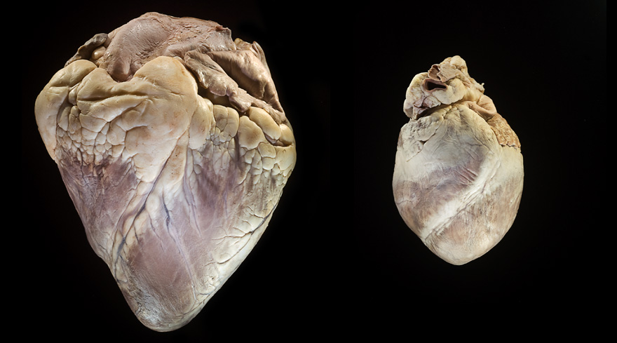
Fig. 1 Bovine heart (left) weighing 4.20 lbs, and Porcine heart (right) weighing 0.62 lbs
About 65,000 heart valve replacements are performed every year in the United States and patients are offered an incredible variety of options for the type of replacement. Many opt for valves made up of biological tissues because unlike mechanical valve replacements, they do not require the patient to take anticoagulants indefinitely to prevent the formation of blood clots on the new valve. Tissues from both pig and cow hearts are commonly successfully transplanted to humans. Figure 1 demonstrates the immense size difference between porcine and bovine hearts. A porcine heart more closely approximates the size of the human heart and whole valves can be transplanted after chemical treatment to prevent rejection. For a bovine transplant tissue from the pericardial sac surrounding the heart is collected and processed into the correct size and shape for the recipient.
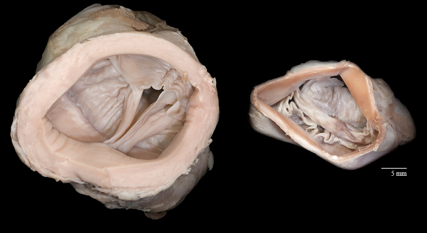
The two most commonly replaced valves are in the more muscular left side of the heart which pumps blood to the entire body. The mitral valve sits between the left atrium and ventricle and is the only bicuspid, or two-leaved valve in the heart. In figure 2, the valve, chordae tendineae (or heart strings) and the papillary muscles, which control the opening of the valve, are pictured. As the lower chamber contracts to push blood out of the heart, the mitral valve is forced closed to prevent the backflow of blood into the atrium. Regurgitation (or insufficiency) occurs when the valve does not close tightly and some of the blood leaks backwards, often due to a congenital defect or Endocarditis. To compensate for not all of the blood leaving the heart, it pushes more blood through, causing an irregular and chaotic rhythm known as atrial fibrillation, which can cause blood clots. Additionally the increased blood pressure in the heart causes further wear on the leaves of the valve, worsening the condition.
Mitral valve stenosis, a condition where the valve does not fully open and less blood is allowed to exit the chamber, often presents about twenty years after an episode of rheumatic fever, which scars the cusps of the valve. Left untreated this causes an enlargement of the atrium as it tries to force more blood through, eventually weakening the heart leading to heart failure.
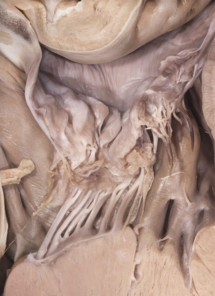
Fig. 2 Mitral valve in a halved porcine heart
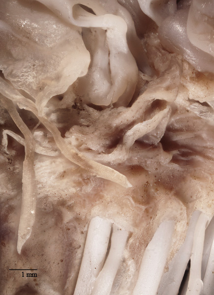
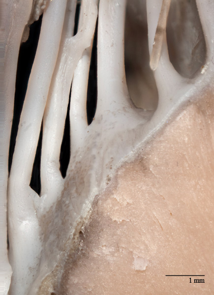
Fig.3 Chordae tendineae attach to mitral valve, 2x magnified
Fig. 4 Chordae tendineae attach to papillary muscle, 3x magnified
The other most frequently replaced valve is the aortic valve, which sits at the top of the left ventricle where it opens into the aorta. The aortic valve is more prone to stenosis. A congenital defect occurring in about two percent of the population causes it to form with only two leaves. Childhood rheumatic fever often also causes scarring in the aortic valve creating a condition very similar to the stiffness caused by calcium deposits in the elderly. All of these conditions cause the blood flow out of the heart to be very turbulent rather than flowing smoothly, causing additional wear on the valve which will eventually require it to be replaced.
Figure 5 gives a unique view of the three cusps of the valve in a preserved bovine heart. An endoscope was inserted into the aorta to image the valve without altering its shape through extraction.
Unlike the mitral valve the aortic valve is not attached to the muscle by chordae tendineae and is controlled completely by the pressure of the blood in the heart. The inferior surfaces of the cusps are smooth as shown in figure 6.
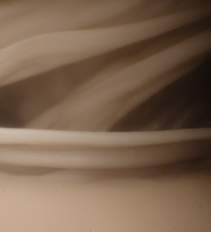
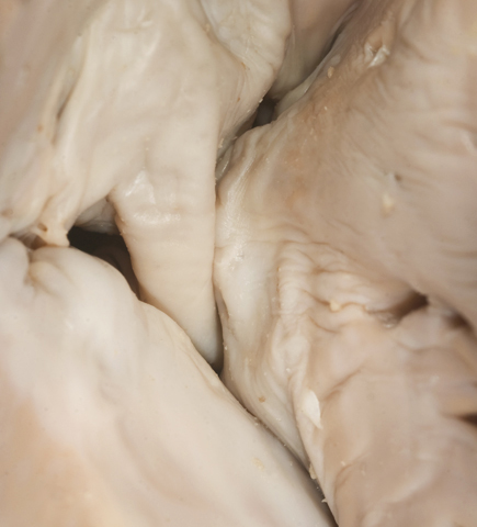
Fig. 5 Endoscopic view of the cusps of the aortic valve
Fig 6. The underside of the cusps of the aortic valve
Both bovine and porcine valves are expected to last between ten and fifteen years, compared to about twenty-five to thirty years with a mechanical valve replacement, so they are a more popular choice among elderly patients who are less likely to have to have another replacement surgery when the transplanted valve wears out. The processing of porcine valves takes about four weeks, during which the valve structure is trimmed and fixed on a flexible frame to be fitted to the patient's valve opening.
As demonstrated in figure 7, a bovine valve is far too large to be transplanted into a human heart even with copious amounts of trimming. Tissue from the protective pericardial sac around the heart is harvested, chemically treated, and ready for transplant in about thirty days. Because of the tough nature of the pericardium, bovine transplants have been shown to last slightly longer than porcine valves, some in excess of twenty years.
As demonstrated in figure 7, a bovine valve is far too large to be transplanted into a human heart even with copious amounts of trimming. Tissue from the protective pericardial sac around the heart is harvested, chemically treated, and ready for transplant in about thirty days. Because of the tough nature of the pericardium, bovine transplants have been shown to last slightly longer than porcine valves, some in excess of twenty years.
Fig. 7 A bovine aortic valve (left) compared to a porcine aortic valve (right), 1/3 life size at capture
| Equipment Used: | Kaitlin R. Corbin |
|---|---|
| Nikon d300 | Rochester Institute of Technology, 2012 |
| 60mm macro lens | Biomedical Photographic Communications |
| Bellows | krc7625@g.rit.edu |
| 38mm thimble lens | |
| Fiber optic lights |
| Sources: |
|---|
| http://www.clevelandclinicmeded.com/medicalpubs/diseasemanagement/cardiology/mitral-valve-disease/ |
| http://www.mayoclinic.com/health/mitral-valve-regurgitation/DS00421/DSECTION=complications |
| http://www.cigna.com/healthinfo/uf4556abc.html#uf4556abc-sec |
| http://www.heart-valve-surgery.com/biological-heart-valve-replacement.php |
| http://www.americanheart.org/presenter.jhtml?identifier=4598 |