Heart Disease is the number one cause of death among men and women in the United States. Statistically, every 25 seconds an American will have a coronary event. About every minute one will die from a coronary event.
The most common heart disorder in the United States is coronary heart disease, which often appears as a heart attack. In 2009, an estimated 785,000 Americans had a new coronary attack, and it is predicted that 470,000 will have a recurrent attack.
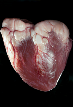
Coronary heart disease (CHD) is defined as a narrowing of the small blood vessels that supply blood and oxygen to the heart. CHD is also referred to as coronary artery disease. Other forms of heart disease are associated with the valves of the heart, such as: Aortic insufficiency, Aortic Stenosis, Mitral Regurgitation – acute, Mitral Regurgitation – chronic, Mitral Stenosis, Mitral valve prolepses, Pulmonary valve stenosis, Tricuspid regurgitation. Many of these are treated with surgery. A heart valve surgery is a delicate procedure that is used to repair or replace diseased heart valves and vessels. For such surgeries a biological valve is often used. Tissues for the procedure are harvested from a human cadaver or from another large mammal. Because of the structural and functional similarities a pig or cow heart is used. Both a cow and human heart have a four-chamber construction, which makes cows a fit donors. One draw back to using a cow heart is the weight of the organ and the volume, but the function is quite the same. The weight of a human heart is between 200 and 425 grams (7 and 15 ounces), and a cow is around 5lbs (2267 grams.) The average heart beat rate however is almost the same (70 and 80 beats a minute) that makes it a near ideal match.
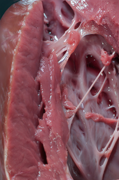
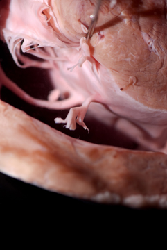
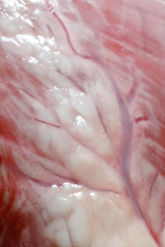
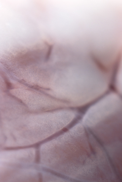
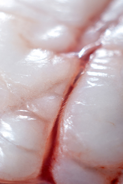
Following images will highlight anatomy of a cow heart
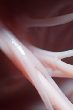
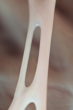
The image above shows the interior of the Right Ventricle. Attached to the heart wall one can observe the chorodae tendineae and the papillary muscle. Chordae tendineae: Thread-like bands of fibrous tissue which attach on one end to the edges of the tricuspid and mitral valves of the heart and on the other end to the papillary muscles, small muscles within the heart that serve to anchor the valves.
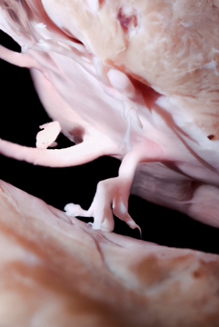
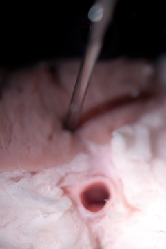
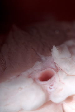
The above image shows the opening of a pulmonary vein. Pulmonary vein: One of four vessels that carry aerated blood from the lungs to the left atrium of the heart. (The four are the right and left superior and inferior pulmonary veins). The pulmonary veins are the only veins that carry bright red oxygenated blood.
Contact Nina Reznik email nxr4674@rit.edu