|
The Virtues of the Diffusion Screen in the 35mm Slide Copier/Film Scanner And its Parallels in Brightfield Microscopy By Paul James |
The practice of siting a diffusion screen at the front of the slide 35mm copier I'd imagined was to ensure the copying device's successful imaging in most of the usual lighting situations that photographers would have at their disposal, for it conveniently broadens the illuminant's field considerably by default and dispenses with the need to rely on purpose made illumination units that would only increase costs. Despite its simplicity it works very effectively. The only proviso being that the colour correction/white balance is taken into account when capturing the image on the digital camera's sensor from any one of a variety of light sources that users might take advantage of.
Fortuitously, the diffusion screen in the 35mm copier not only broadens the field of illumination sufficiently to flood the entire 35mm format......its principal purpose, but it also manages to severely reduce any high contrast diffraction which would have been raised in profusion by all the hard edged unwanted particulates and markings that might litter or are embedded in an old film emulsion. Conducting an A-B test with and without the screen in place soon reveals its effectiveness in this situation, though the relative differences between the 2 types of illumination depends upon the alternative light source used besides the diffusion screen. A near parallel light source will reveal every speck across the emulsion and film base with great clarity. Of course the more robust particles and marks cannot be drowned out from the image by diffused lighting, but the greater proportion of the unwanted finer dross can be dispensed with, which is remarkable given the utter simplicity of the situation.
The All Important Illumination........Taking the technique to extremes.
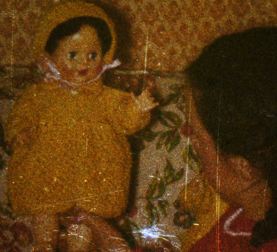
I thought I'd show as a classic example of how the process of imaging by 2 entirely different lighting techniques can produce 2 radically different effects. An enlarged crop ( Photomacroscope) from a Kodachrome colour slide taken in 1954 which is in poor condition is first shown illuminated with partially oblique lighting ( ie a near parallel source tilted to one side ). The depiction of detail is severely emphasised, especially of grain structure, particulates and other markings such as scratches are highlighted very effectively. Diffraction artifacts and colour distortion and entirely unsuitable in this situation :-
|
|
|
|
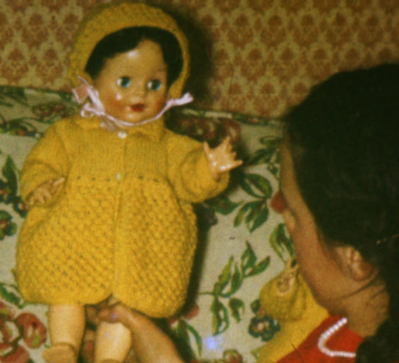
The second image above was raised by near parallel lighting in line with the optical axis of the macroscope but with a plastic diffuser in contact with its emulsion layer. Thus the light rays emerging through the diffuser would be randomly deviated through a very wide angle approaching 180 degrees, though the bulk would be less than this. Note the virtual absence of markings and fine debris across the emulsion. Despite the severity of flooding the film emulsion with chaotically disorganised diffused light, the image remains intact and visually comprehensible.
The abundance of 'detail' in the first image is overly exaggerated by oblique lighting, and though it is a most useful lighting technique in microscopy for that very reason, its ruthless exposure of every nuance of detail simply spoils the more subjective elements of the image. The opposite extreme shown in the diffuser image efficiently rids the subject of unnecessary distractions of fine detail induced diffraction rather dramatically, though it can't remove major defects near centre bottom and others of less importance. Despite the diffuser's apparent detail and contrast moderating properties on film emulsions, the overall effect is very satisfying especially at normal viewing scales.
B&W Negative Treatment
|
|
|
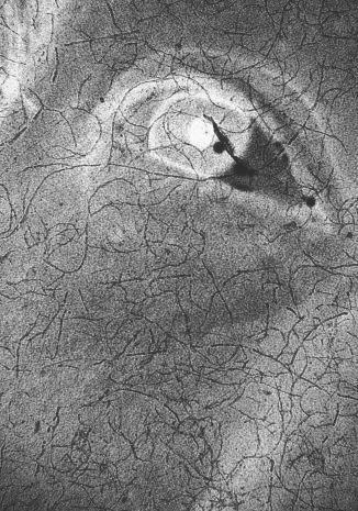
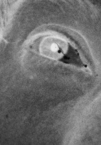
Above left is shown a small crop of a B&W negative which is about 45 years old, with fungal hyphae infestation. It was illuminated by near parallel light only. The image right taken with the diffuser screen was in contact with the emulsion using the same illuminant. Again a dramatic reduction of unwanted artifacts. The grain's acutance seems to have been slightly muted too, but I think this can be a positive benefit in the presentation of portraiture.
The image left appears crisp in comparison to that shown on the right. But it must be remembered that a large proportion of the 'detail' in the image shown left is actually composed of diffractive anomalies from both fungal hyphae as well as the silver grain particles. Thus the diffraction bereft image right appears soft by comparison. The underlying image remains similar in both examples.
These intimate views of the film's surface were imaged through the photomacroscope. I used it because its slightly higher amplification of the film reveals the differences more easily onscreen. The bright field illumination was generated from a single LED suitably modified for macro fields by a lamphouse condenser, and behaved as a near parallel source. The images are straight out of the camera ( Sony NEX 3 body ) and cropped for convenient onscreen illustration.
In the darkroom situation, these imperfect negatives respond to a diffuser screen in the enlarger in similar fashion.
Parallels In Bright Field Compound Microscope Practice
The reality of what is going on within the slide copier is loosely comparable with the illumination techniques of Bright Field microscopy. The observation of a semi-transparent specimen such as a film negative/positive whilst opening and closing the substage condenser's iris diaphragm will reveal a similar range of suppression of diffractionally orientated detailing. The wider the condenser's aperture the greater the suppression of the specimen's semi-transparent structure : Conversely as the iris closes down, the pencil beam passing through the specimen is much nearer a parallel beam and exacerbates the presence of diffraction patterns of minute particulate or linear form greatly. The skill in brightfield microscopy is in developing an awareness of the importance of the setting of the substage condenser's iris diaphragm for a given specimen. Yet despite this well understood imaging principle, the use of diffused lighting for certain subjects that are of a more robust nature such as insect parts etc., greatly enhances an otherwise overly contrasty image that conventional brightfield tends to raise in this situation. The diffuser can, if suitably close to the under surface of the slide generate moderately high NA light and obliterate virtually all the diffractive components in one process. So a fair amount of potential resolution is maintained, but contrast is muted, but not too severely. Since the colour slide/B&W negative's base emulsion detail is more robust than many a microscope specimen's delicate semi transparent structure, the muting of contrast that diffusers can cause does not change the image information to any significant degree.
Proximity of the Diffusing Screen in the Slide Copier
The diffusing screen's efficiency at removing unwanted dross and markings is at its greatest when in virtual contact with the film's emulsion. But this requires that the diffuser has no base granularity such as that found in ground glass, since this matrix will be captured in the image. A pity since ground glass has little or no effect on colour balance that some of the other diffusing screens seem to have. Flashed opal diffusers are very good though costly, but they can, like the plastic diffusers be placed against the emulsion side if necessary for maximum effect when imaging the very worst examples of film in poor condition. Too intimate a contact can raise Newton Rings which will be counterproductive. Yet even at a distance of about 10-20mm from the emulsion plane, where the plastic diffuser in slide copiers is commonly sited, the suppression of unwanted artifacts is still satisfyingly efficient.
In the worst case scenario of film condition, it maybe necessary to employ a wet mounting of the film between glass slides. This can nullify the major marking/scratches by cancellation of refraction that will occur in the angular interfacing in these grooves in the acetate film base. It is a messy process and not easily contrived to remain bubble free for the duration. However for 99% of our aging nostalgic slide and B&W negative collections, the dry diffusion screen will no doubt make a significant difference to their presentation. In the final analysis personal preferences reign regardless.
Diffusion Screens and The Film Scanner
The effects of diffused light extend to the 35mm dedicated film scanner too as illustrated below. Again unsized crops from B&W film around 45 years old, but scanned in a Minolta Dual Scan 111. A flashed Opal diffuser was used here, placed against the film's emulsion layer where the effects of Newton Ring generation were considerably reduced compared to the potential generation of NR raised if the opal glass was positioned against the film's glossy base layer. Again the differences between the images are clearly obvious. If any softening of the image has occurred by the process then it is very subtle and from my perspective welcome in reducing the coarseness of the silver grain very slightly. It appears too that the image's contrast has been somewhat reduced :-
|
|
|
|
The 'cleansed' appearance of the resulting image can fool the subconscious mind into believing that there has been a softening of detail simply because the acuity of the grain structure is somewhat reduced along with the removal of fine scratches and the like. Conversely, from another perspective, the presence of the sharp representations of the unwanted detritus on the normal non diffused lit image can trick the subconscious into extolling the virtues of the original taking lens, which of course played no part in their appearance ! I think there is much psychology involved in the appraising of imagery.
Last Word
The perceived improvements from this simple diffuser technique are elevated in part, by the entirely subjective viewpoint we all hold concerning imagery, and most especially of the human face. I suspect a favourite photograph is what we want it to be, and little else. The humble diffusion screen, often maligned in microscopy, helps in part to achieve just that.
| All comments welcome by the author Paul James |