A Review of the Quality and Usefulness of Anatomical Models
by Mol Smith Nov. 2013
Introduction
Microscopy and Biology are related. In fact, I would say they are joined at the hip... if you excuse my weak pun. You may study the biological processes of us humans and other living forms with your microscope, or teach others to do like-wise, but you still need to relate those slivers on your slides with flesh and blood people. It's quite difficult for most of us to slice up a willing subject and take a look inside at those vital organs. Thankfully, we can resort to Anatomical models instead, which is a lot
less messier and traumatic.
I wrote to Instruments Direct (Services) Ltd in the UK to ask if I might borrow one of their models to review. You see a lot of these on Chinese web sites but I wanted to see one which promoted itself as high quality and well made, which is what IDS models appear to be from their
web site page here. They were very kind and - without asking for any bias to be given (such is their positive confidence in their products) - sent me a model
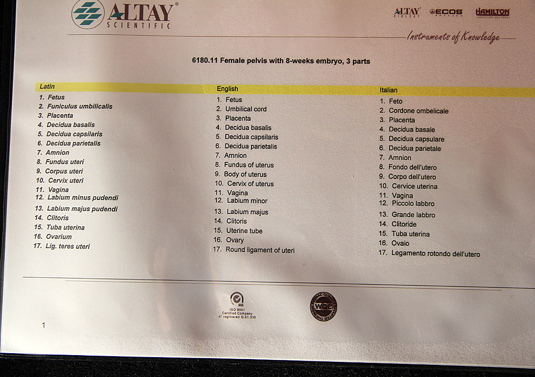
of a Female Pelvis with 8 week Embryo.
Packaging
Well packaged with sufficient protective bubble wrap, protective paper on the model (non-sticky) and arrived efficiently via recorded delivery in a robust cardboard box. No problems or worries about receiving something knocked about by Mr. Postie here, so we're off too a good start.
Let's take out first look at the model so we can see what we're talking about.
Three parts
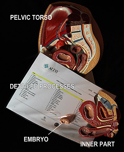
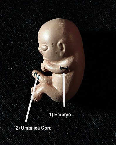
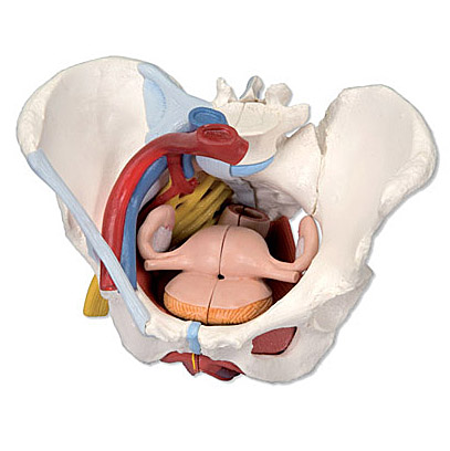
The model is composed of three robust plastic parts: the pelvic Torso, partly cut away, the inner Torso - removable, and a tiny fetus (foetus) resting in the womb. Three sturdy metal pins hold the second largest part to the main body via accurately aligned locating holes. (See inserts below).
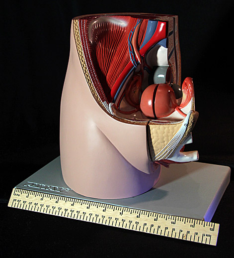
| Out of the box and unwrapped. The base is 12 inches across. Lifelike colours. Sturdy. Robust. |
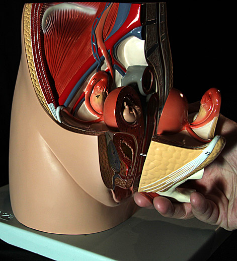
| The second largest part pulled so the locating pins exit their holes, revealing the embryo. |
| Everything. The model in 3 parts, and the very detailed processes card, numbered. |
The weight of this immediately tells you it's not a cheap low-quality item. It feels good and it looks great. The colours of each of the biological processes is well done aiding quick identification or various organs and internal anatomy. The painting or colouring of the plastic parts looks well done and not likely to wear off easily. The base has 5 screws through from the underside into the model, allowing optional detachment from the base.
Other than flat diagrams and visual 3D modelling, this is the first opportunity I had to look inside the female body. Any body in fact! I thought I would know more but it's surprising when looking at a physical 3D model how different it presents itself compared to your previous experiences. What was crucial and worked well, helping me to learn how the reproductive system worked and how it remained separated from the waste discharge system, was the attention to detail in numbering all the different parts. A quick look at the reference guide (a laminated 2 sided sheet), and I was rapidly identifying everything and their relationships to the other processes.
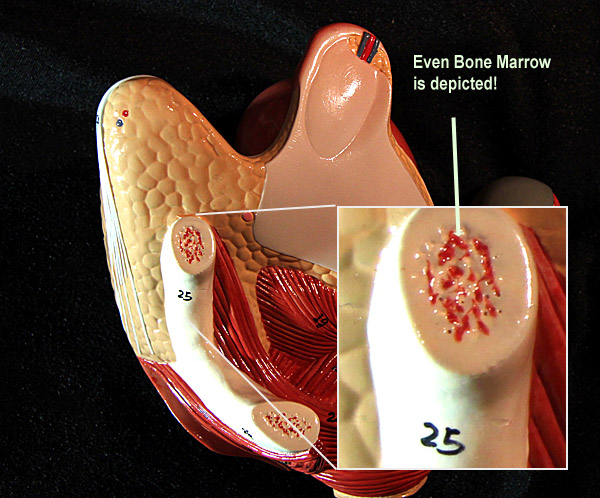
The Devil is in the Details

The model stands up to close scrutiny. Internal parts are properly scaled and sculptured. Tiny hand painted numbers mark each process in a refined and detailed manner, even the umbilical chord running from the foetus. |
The images below show the attention to detail and referencing. It is fairly obvious that these models are NOT made in China as their quality and workmanship suggest European craftsmen. |
|
 |
 |
Priced at just: Ł85.00, this model is exceptional quality for its low price and although I am not here to sell products, I can only recommend it highly as a invaluable teaching aid. I was so impressed, I went to the web site to see the huge range of anatomical models sold by IDS. Here are a few samples from their wide and varied range. Many models are hand painted in Germany. All of them live up to the same high quality in detail and importance for Anatomical Education.
 |
 |
 |
|
Female Pelvis With Ligaments, Vessels, Nerves and Pelvic Floor Organs. This life size six part model of a female pelvis represents detailed information about the topography of bones, ligaments, vessels, nerves, pelvic floor muscles and female genital organs. It presents the whole pelvic floor with partially removable midsagitally sectioned external anal sphincter, external urethral sphincter, deep and superficial transverse perineal and bulbospongiosus. |
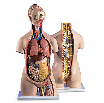
Classic Uni Sex Torso. 18 Parts Open Back. This classic torso is hand-painted true to detail and made of high-quality plastic. All 3B torsos are also developed and modeled in Germany. This human unisex torso model has the unique feature of an open neck and back section going from the cerebellum to the coccyx. Vertebrae, intervertebral discs, spinal cord, spinal nerves, vertebral arteries, and many other features are represented in detail in this colorful replica of the human anatomy. Price: Ł373.00 |
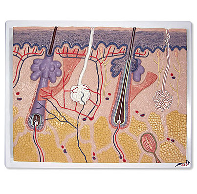
Skin Section. 70X Lifesize. This skin relief model shows a section through the three layers of the hair-covered skin of the head. The skin section shows: •Representation of hair follicles with sebaceous glands•Sweat glands•Receptors•Nerves•Vessels Price: Ł55.00 |
This is only a small sample from an extensive range. I'm not here to sell their products, and to be honest, I wanted to see how good some of these models are. But, I am quite staggered by their high quality, reasonable cost and by the advantages of being able to touch and feel physical models to fully understand them. I would highly recommend these models sold by this company to all educational institutes and learning centres.
Please note: photos are copyright. Please request permission before using.
References:Models are sold by: Instruments Direct (Services) Ltd
Unit 8 The Courtyard, Stenson Road, Coalville, Leicestershire. LE67 4JP. UK.
Tel: +44 (0) 1530 832500 Fax: +44 (0) 1530 817087.
Email: sales@inds.co.uk Web Site: www.inds.co.uk