A Pier Without Peer
Richard L. Howey
First of all, I want to thank my wonderful and generous friend who sent me that sample I am going to talk about here. Mike Shappell and I shared many microscopic adventures when he lived here in Wyoming and now he and his wife Kelly share the wonders of nature and microscopy with their three teenage children. They lived for a number of years out in a semi-rural area of Washington State near a lake where wildlife was abundant. Now, however, they are all living in the world’s largest city, Tokyo, but are still able to find areas to explore wildlife including the sea. Some time ago, while still living in Washington, he send me a plastic container of material which he had collected from a piling on a pier in Puget Sound (how’s that for alliteration?) It was nicely preserved in alcohol and is just the sort of random concatenation of organisms that thrills and rejuvenates my ancient mind and body.
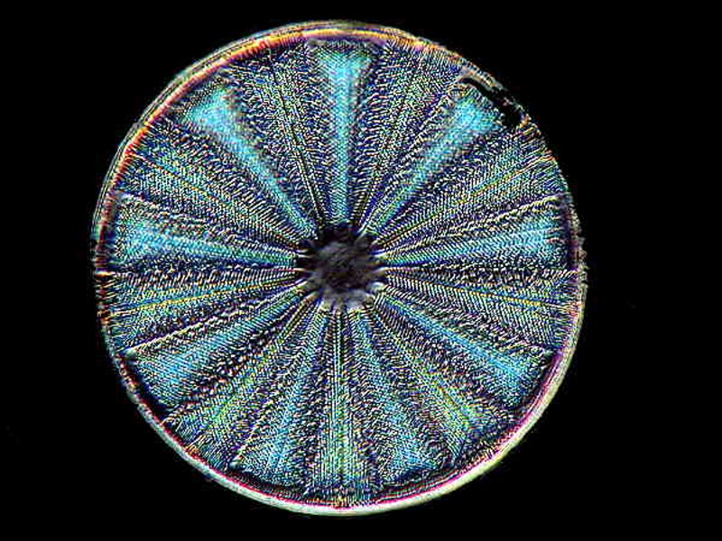
I have an eccentric procedure for investigating such samples. My first priority is the detritus in the bottom. I pipet a modest quantity of it into a small Petri dish and then begin to scan at 20 to 40 magnifications and if I find something especially interesting, I’ll zoom up to 80x. The first thing that caught my attention with the first dish was the diatoms. Diatoms, as most of your know are extraordinary organisms which extract silica from the water and build glass houses consisting of two overlapping frustules forming a valve. We humans complain about living in glass houses; how frustrating it must be to live in a pair of frustules! Diatoms are some of the most elegant and extraordinary items to be found on Planet Earth. Hey are generally divided into two major types: he centric diatoms and the pennate diatoms. The circular diatoms such as Arachnoidiscus are readily identifiable as centric, but there are other forms that are triangles, squares, or pentate (5-sided) which are also regarded as centric.
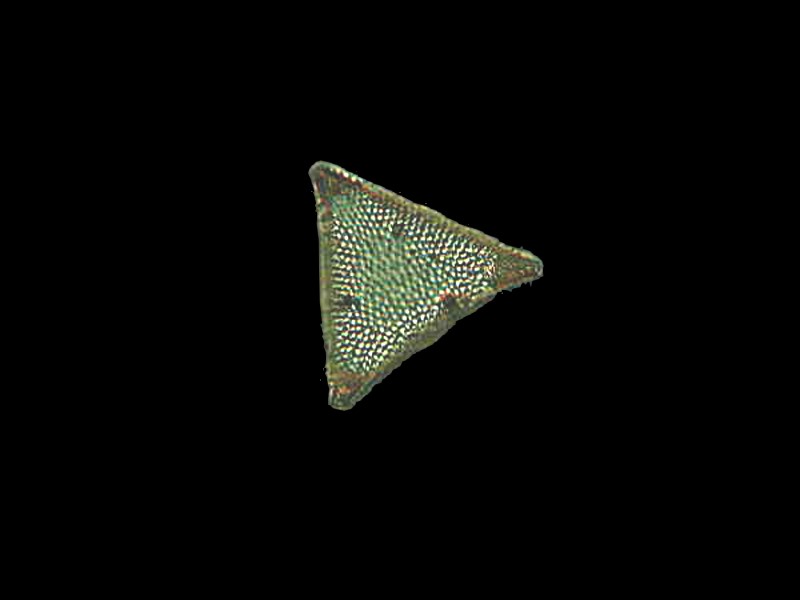
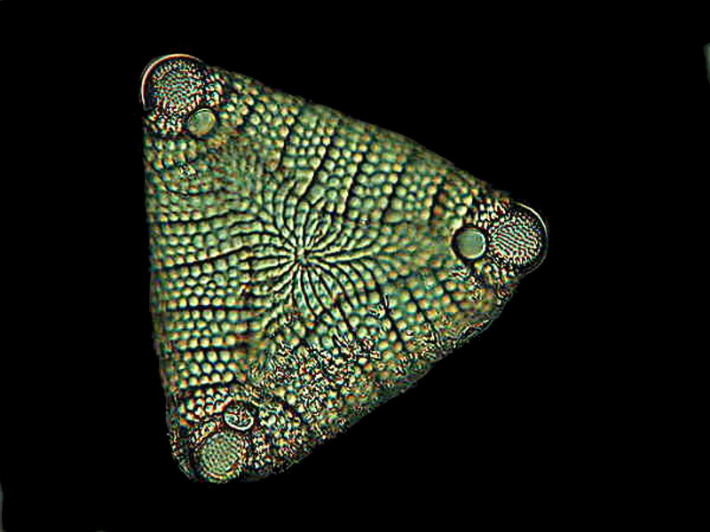
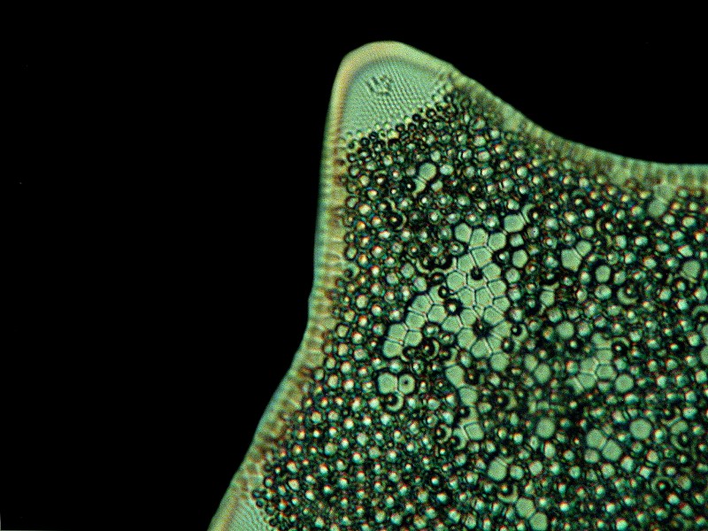
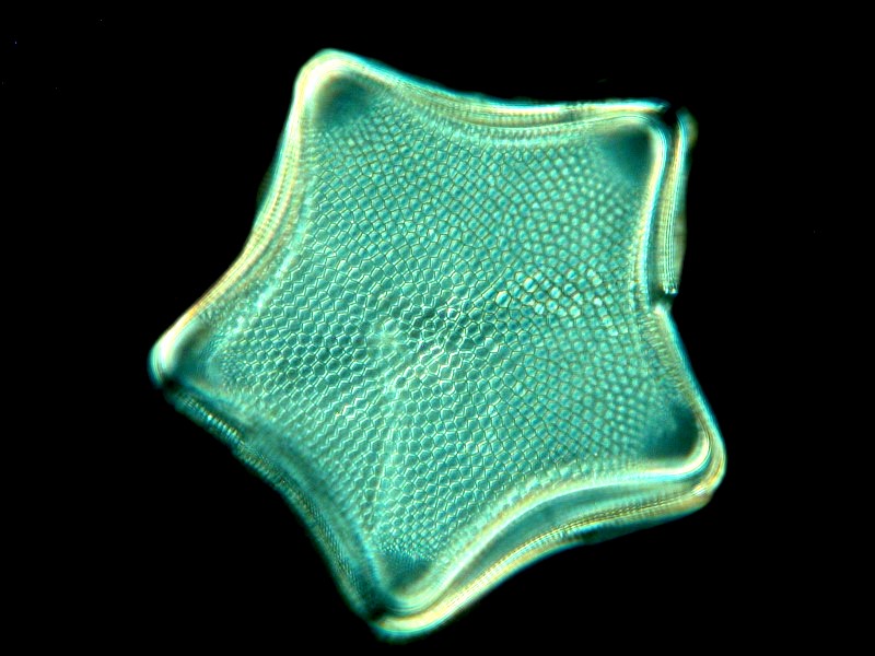
The justification for this is that the “dots” or pores converge on the center which you can see when you look at the images closely.
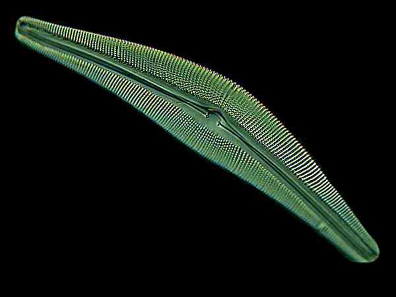
The pennate diatoms are often long and relatively narrow; some are shaped like miniature canoes, others like small thin rulers, and yet others are almost needle-like.
Here is an example of Cymbella.
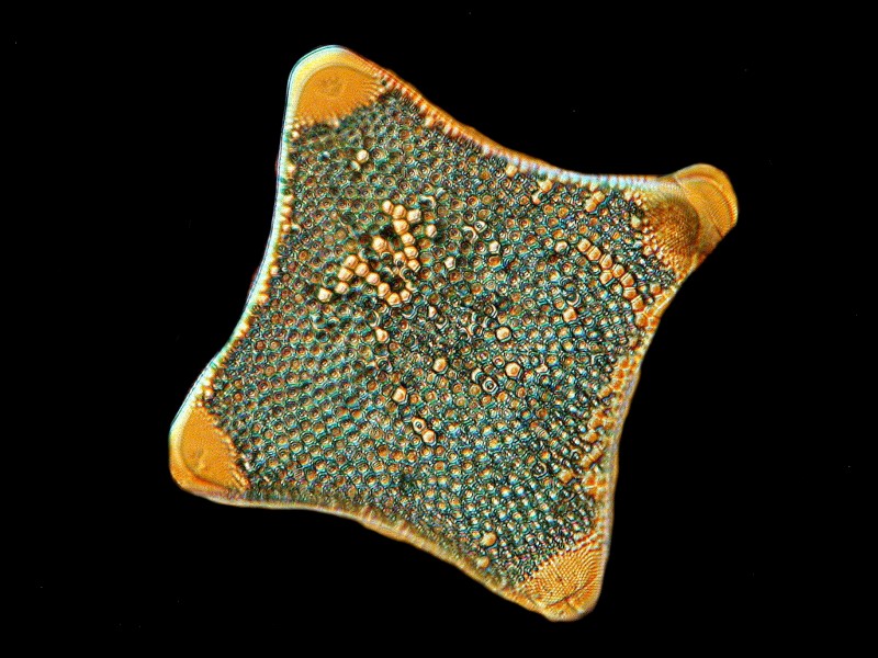
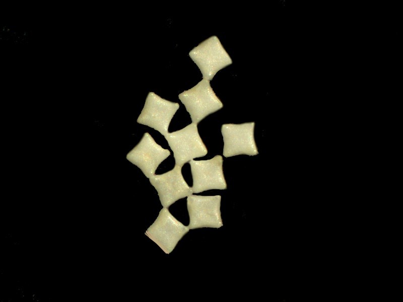
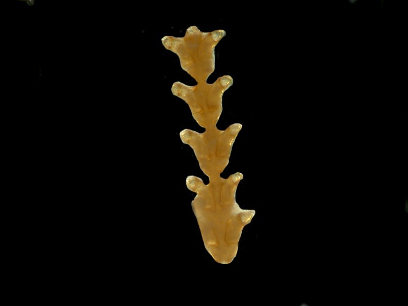
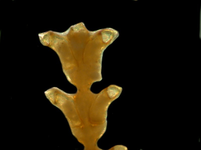
To make things even more interesting, there are some stunning species that form colonies or, at least, aggregations. The square ones which I found in this sample occur singly, but also in clusters of anywhere from 2 to 16 (which is the largest grouping I found). They connect at the corners as you see from the image below.
Of one of these forms which aggregate, Wim van Egmond has produced a superb image of Lichmophora fabellata and it looks like an exotic fan.
And there is a species called Diatoma vulgaris which looks like an old-fashioned carpenter’s rule partially unfolded.
For those of you who really want to get up close and intimate with diatoms, there is a splendid book with 2,500 Scanning Electron photomicrographs titled The Diatoms: Biology and Morphology of the Genera by F.E. Round, R.M. Crawford, and D.G. Mann. Unfortunately, the hardback edition which has been out for some years runs about $300 on a good day. However, for those of you who are willing to make an investment, there is some moderately good news–a very nice paperback edition is now available for $80. If this is still too much, blackmail your local librarian. It really is a superb reference book. Diatomists of the 19th and early 20th Centuries, were, I think, a bit like the Mad Hatter in Alice in Wonderland. However, instead of being made mad by the mercury used in tanning, some of the diatomists risked using arsenic compounds or boiling concentrated acids, such as nitric acid, the vapors of which will not only clear your sinuses, but dissolve your brain. I could go on and on about diatoms, so I recognize that it’s time to move on since Dave Walker, generous though he is, has limited me to a petty 8 million words for this essay.
What other delights are to be found in the detritus of this sample? As one would expect, there are various kinds of eggs and generally speaking (99% of the time), it is pointless to try to identify them. I found a foram shell or two; a number of nematode “worms” with needle-sharp proboscises, proboscisuses, proboscii (or whatever)–needle-sharp feeding tube; ostracod shells; copepods; minute polychaetes, many small crustacea; hydroids, bryozoan bits; bits of algae, some of them are delicate red fronds which have retained a fair portion of their color. This tempts me to launch off on a long digression about the biological significance of color with an excursus on why some preserved specimens tenaciously preserve their pigmentation whereas others lose it very quickly when preserved. However, I shall spare you such a long-winded discussion until I write another essay devoted to that topic.
On the largest scale organisms (about 3 inches), we find several tunicates with lots of things attached to them. One of the tunicates is of the genus Styela with its typical “warty” surface and distinctive siphons. There is another genus which has a very tough tunic and when I cut through the area of one of the siphons to extract and study it, I discovered a bright orange tubular structure extending downward from the siphon. This one is completely new to me and requires further investigation.’
On the surface of one tunicate, I found a small (2 inch) gelatinous, nearly transparent mass which is yet another tunicate, a colonial one. I extracted one of the members of the colony and could see the 2 tiny siphons and the lacy network of slits in the pharyngeal basket.
On the underside of one clump, I found a sand-coated tube about an inch long which had a small section missing and there in that opening were some tiny opalescent bristles–a clear indicator that we very likely have a polychaete worm. I carefully removed the tube from its embedded surroundings and discovered at one end, a bundle of tentacles. Then, at the bottom of the container, I noticed another feathery sort of object which turned out to be a 2 inch long polychaete, but the second best part was that the tentacles were nicely extended and the first best part was that both specimens had retained a bit a pigment in the tentacles and were, in fact, what is popularly know as feather duster worms.
This afternoon, I scraped the surface of a Styela –algae, centric diatoms, pennate diatoms, colonial diatoms, humpback whales (just checking to see if you’re paying attention), bits of hydroids, and several species of sponges. Then the next stage is to remove the largely denuded Styela and place it back in the alcohol for later further investigation.
At this point, we can poke yet again through the scrapings and see what else might show up. Almost immediately, two objects of considerable interest to me came into view–both of them larval forms. Larvae present an enormous challenge when it comes to identification. Many of the early stages morphologically bear little resemblance to their later adult forms. Fortunately for us, there are researchers with enormous patience who have meticulously studied the development of a wide variety of extraordinarily challenging organisms and, in my view, this is especially true with regard to the invertebrates (politicians, university administrators, civil servants, etc.). Embryology and developmental biology require special facilities, remarkable dedication, and the ability to see highly subtle connections. Certain starfish and sea urchins have been the objects of such investigations in an intensive and extensive way. Time permitting, I’ll return to this subject a bit later.
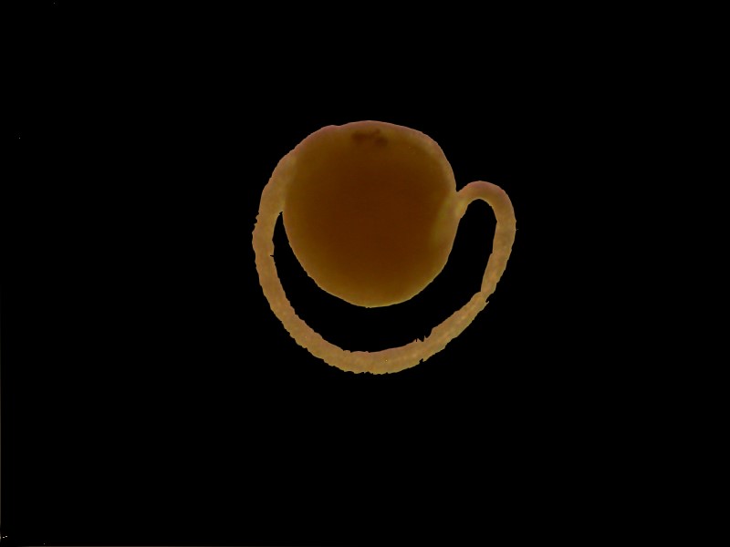
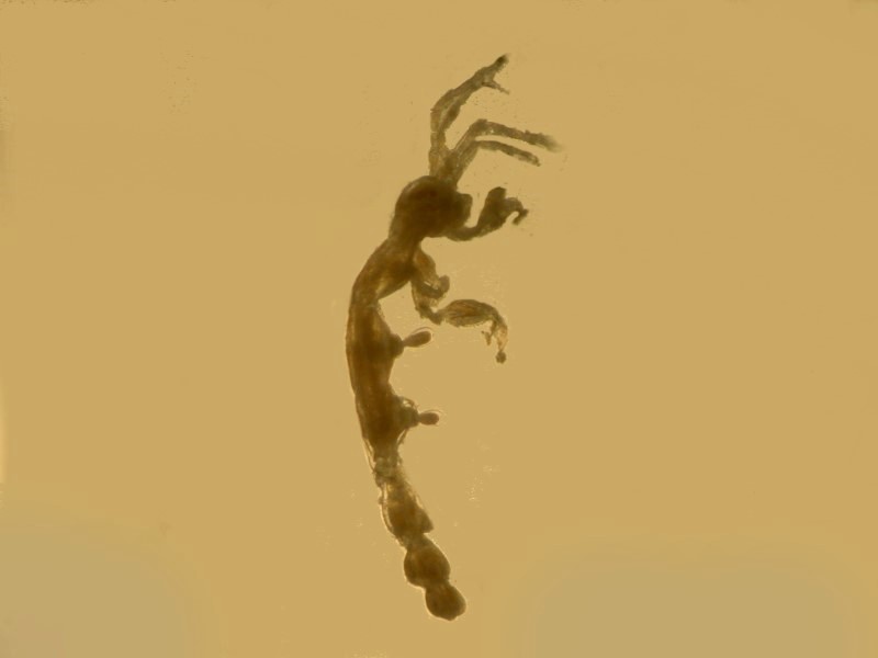
The first larval form which I found is, at this point, still a complete mystery to me.
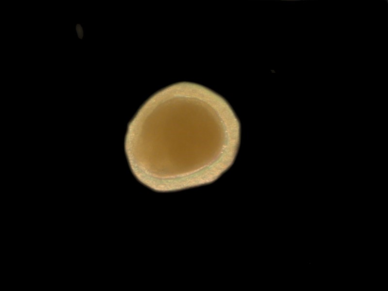
However, at first, I thought that this was an egg until I found a second specimen and then it became clear that it is a larval form of some sort. As you can see, what appears to be a surrounding membrane in the first image is in fact a tail.
However, some of those dedicated researchers have produced enormously helpful reference books for those of us amateurs who are either too lazy, too busy, or too poor to be able to conduct such studies. I will mention a book which I have found very helpful. It is a large format paperback of 626 pages called Atlas of Marine Invertebrate Larvae, edited by Craig M. Young published by Elsevier which means that it is outrageously expensive. It was originally given to me as a gift just shortly after it came out and it was somewhat over $100, but now a new copy is $1751.92 and a used copy is $656 !!! Whenever I find a highly technical reference that I know will appear in a limited edition, if it is at a reasonably unreasonable price when it first appears, I buy it knowing that it will inflate to an unconscionably excessive price in a very short time.
Virtually all larvae are planktonic, but some attach very quickly to a substrate or the surface of another larger organism as a strategy to avoid becoming prey. Nonetheless, planktonic roms are often produced in staggering numbers because the rate of predation is so high, thus a single oyster may produce millions of eggs and although the larvae develop in a few hours, they may not attach for several days.
Books that deal with larvae also often deal with planktonic pelagic forms that spend their entire adult lives as free-swimming adults. These include copepods, krill, siphonophores, ctenophores, intriguing worms such as Tomopteris, medusae both large and small, diatoms, desmids, squid, protozoa and coccolithophores which though they rarely exceed 10 microns in size can “bloom” in such enormous numbers that our satellites can get images from space of areas of the ocean where the density of these organisms is so vast that there is a distinctive change in the coloration of the sea at such points.
However, the ones which we are concerned with here are mostly those which are sessile (that is, they settle down and attach to something). It is, of course, necessary to recognize that there are numerous form which spend their lives crawling around on or burrow into the surface of substrates or live on other organisms which, nonetheless, aren’t attached in the sense of being physically anchored and thus have the option of moving from place to place but, strictly speaking, are not pelagic. Caprellid shrimp and nudibranchs can frequently be found crawling around on masses of mosses and hydroids. Nudibranchs (also know as “sea slugs” or as irreverent researchers refer to them “nudies”) are among the most marvelously colored organism in the sea. Check out a series of sites with wonderful images which you can find by Googling “sea slugs”.
What is so splendid about even preserved samples (collected responsibly with proper permits) is that an amateur like myself who lives 1,200 miles from the sea can get a glimpse of the marvelous complexity of an aquatic marine community in which virtually every niche has a sub-niche. Even a small sponge may have tiny polychaetes that have ‘wormed” their way into the protective embrace of the sponge and, if there are any micro-algae growing on the sponge, you have a virtual guarantee of finding diatoms and forams.
Speaking of polychaetes, there was a tunicate “embraced” by several tubeworms in such a fashion that it was impossible to tell which ones were there first. Also of interest is that face that the tubes, though flexible, are extremely tough, almost as leathery as the tunic of the ascidian (tunicate). Two of the tubes are 3 or 4 inches long (when uncoiled), but there was a third small one which readily yielded up its inhabitant. In addition to its tube, it has a transparent membrane which surrounds it and which had swollen sufficiently to clearly exhibit some clusters of setae (bristles).
These are birefringent, as you can see in the image below taken with polarized light.
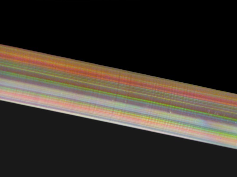
The setae are designed to allow the polychaete to move along inside the tube and up to the anterior opening where it can extend its tentacles to feed. In these specimens, at least, they are located on the ventral side and the dorsal side seems, in its somewhat swollen condition, to possess a series of small transparent sacs. This clearly requires further investigation. Also on this tunicate-polychaete complex are pieces of barnacle, small sponges, and remnants of hydroids.
So now, it’s time to tackle the tunicates themselves which means a bit of discussion about the key characteristics of ascidians. 1) Image a pear-shaped sack with a blunt tip and this is where the 2 siphons are located, the incurrent and the excurrent one. This sack is enclosed almost suspended in a “coat” (mantle or tunic–thus tunicate) which can range from a thin, semi-transparent, membranous “container” to a thick, leathery opaque covering so tough that it requires bone cutters to remove sections for examination. Furthermore, these tunics are largely composed of cellulose compounds, a relative rarity in animal tissues.
2) The sac inside the tunic is rather like cheesecloth in structure and possesses countless cilia which draw in water to be filtered for food particles and the excess water and indigestible waste is then ejected through the excurrent siphon.
3) There is a heart which has 2 “pacemakers”; the heart beats 200 or so strokes in one direction and the reverses while the first pacemaker “recharges”.
4) Some ascidians are individual and others are colonial. Some colonial forms arrange themselves in a circular pattern on a flat substrate producing the appearance of a tiny flower, Botyryllus schlosseri, for example. Others combine to produce large gelatinous masses called “sea pork” by sailors, which can contain thousands of tiny tunicates.
I seriously doubt that these were eaten, but I certainly won’t dogmatically assert that. Some years ago, I wrote an article titled: “Tunicates With Salad On The Side”, in which I brashly asserted that tunicates are not edible. Well, I got a number of e-mails informing me that that is simply not true.
In fact, I learned that in Chile, Peru, a number of southeast Asian countries, and certain island countries, some species of tunicates are regarded as delicacies. I was going to write a followup article addressing this topic when my computer crashed and I lost all of the e-mails which dealt with this issue. However, I do remember a few salient points. Several messages admonished me for not knowing that in certain places, tunicates are regarded as a speciality dish which is highly prized. I do recall an e-mail from a man from Texas who had returned either from Peru or Chile where, at the urging of friends, he had tried tunicate. His description, as best as I can remember it, was that it tasted like and had the texture of a burnt rubber truck tire which had been soaked in iodine. No, it might be, that slices of “sea pork” properly marinated and seasoned could prove delicious but, in spite of my 65 year long fascination with tunicates, I can guarantee that I’m never intentionally going to eat one.
I mentioned find an encrusting colonial tunicate on a piece of kelp. It is a species of Didemnum which often has a whitish, granular appearance inside the enveloping membrane. This is a consequence of tiny, spiky, spherical calcareous spicules which are difficult to isolate because the usual tissue-dissolving techniques don’t work for cellulose compounds. So, at this point, I got out my sharpest micro-scalpels and cut the tiniest sections I could manage. One batch I set aside to work on using just manual micro-dissection techniques. The other batch, I subjected to treatment with a solution of the enzyme cellulase in the hope that it would dissolve out the cellulose compounds. If I get results in time, I’ll let you know here; if not, I’ll write them up for a future essay.
As I said at the beginning, I love poking around in the bottom debris and seeing what goodies are hidden there. To conclude this essay, I’ll give you a small gallery with a few brief comments on some of the critters I have found so far.
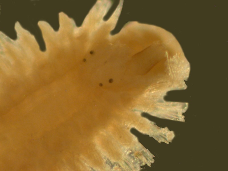
All of these were examined and photographed using an Olympus SZ trinocular Stereo microscope fitted with a 2x objective thus provide a magnification range of 14x to 80x. I used incandescent transmitted illumination, a halogen ring illuminator and/or a flexible fiber optic halogen illuminator for incident light. By using various combinations, I was able to see different sorts of detail with different lighting techniques. For example, here is an image of a section of a polychaete worm using both transmitted an incident light.
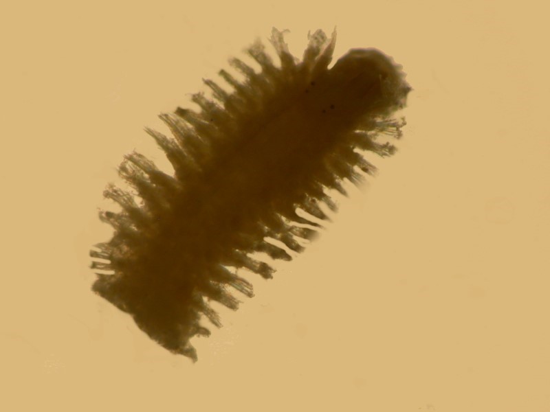
Increasing the magnification reveals that the worm has 4 eyes and is covered with debris.

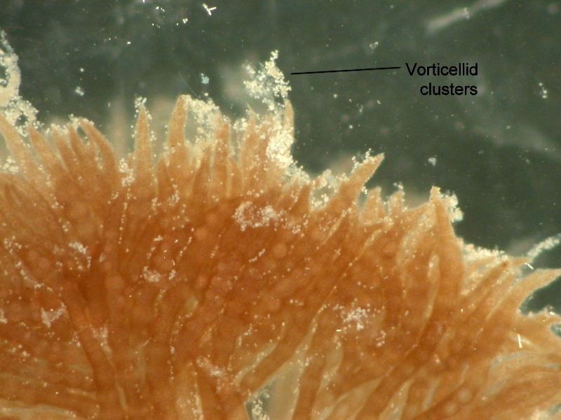
By focusing closely on the setae, I was able to see tiny protozoans (Vorticellids, in this case) which had been preserved there in a contracted form.
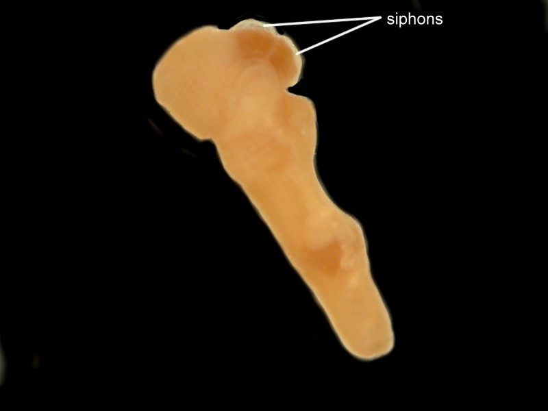
Above, I mentioned colonial tunicates. Well, in the detritus, one can find individual specimens which got dislodged from their colonial matrix. Here is an example.
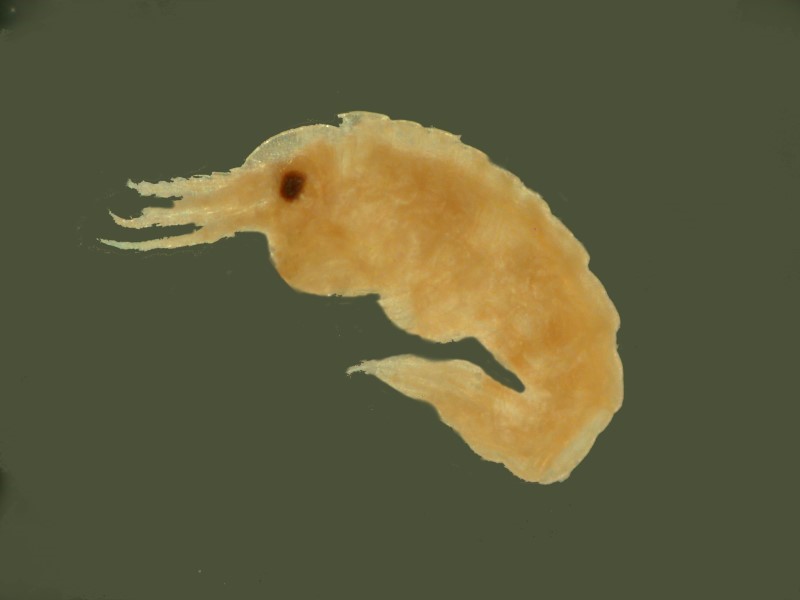
Here, in this strange mixture, one can also find tiny relatives of shrimp; in fact, I found 2 kinds. The first is more traditionally shrimp-like.
The second is a Caprellid or “skeleton shrimp”. By playing with the lighting, I was able to capture some of its rather eerie character.
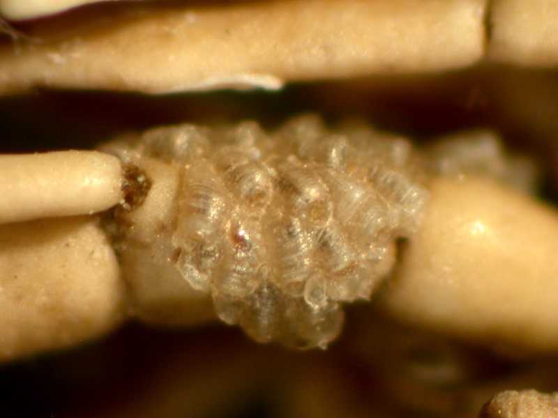
Fragments if bryozoa or “moss animals” are common and I’ll show you three images to underscore the difference that experimenting with the lighting can make.
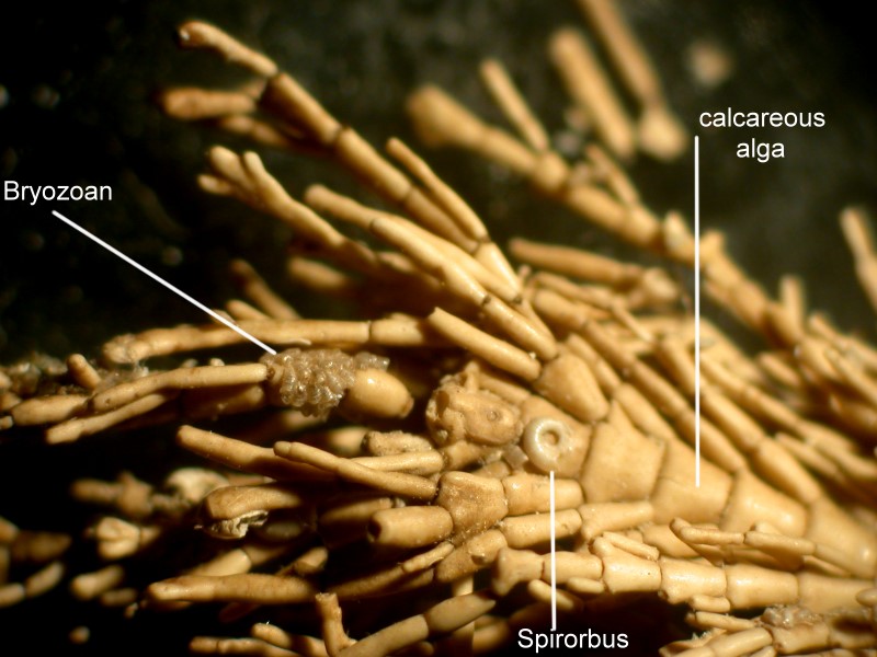
In this sample were some pieces of a calcareous alga which is quite wonderful to investigate. The outer surfaces are coated with carbonate deposits and so give the appearance of being “skeletonized”. On such a piece, I found another kind of bryozoan and also the shell of tiny tube worm (Spirorbis) as well.
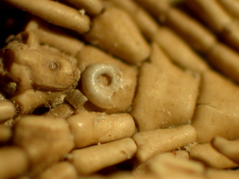
Here is an image showing the calcareous alga, a remains of a small colony of bryozoa, and a damaged shell of a Spirorbis.
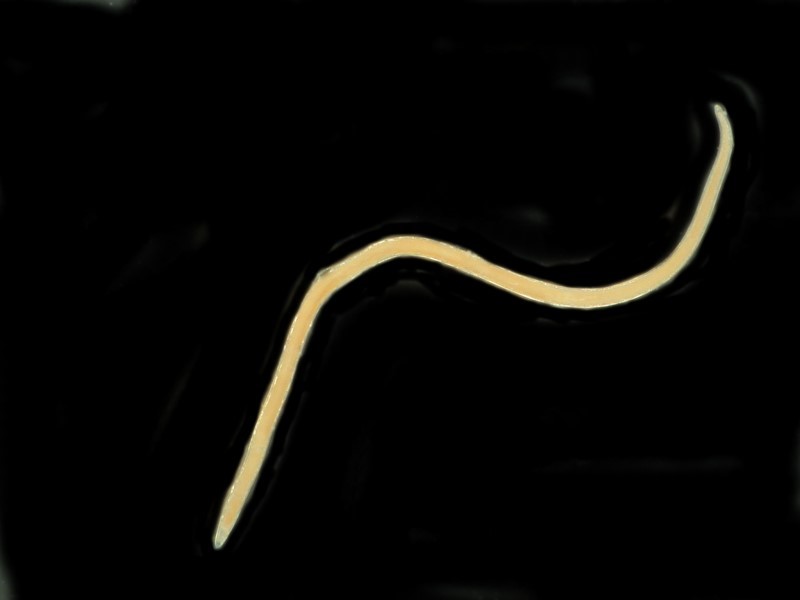
Another sort of “worm” shows up frequently, namely, nematodes.
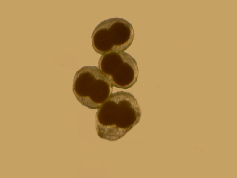
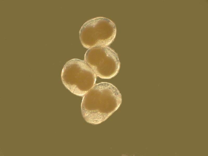
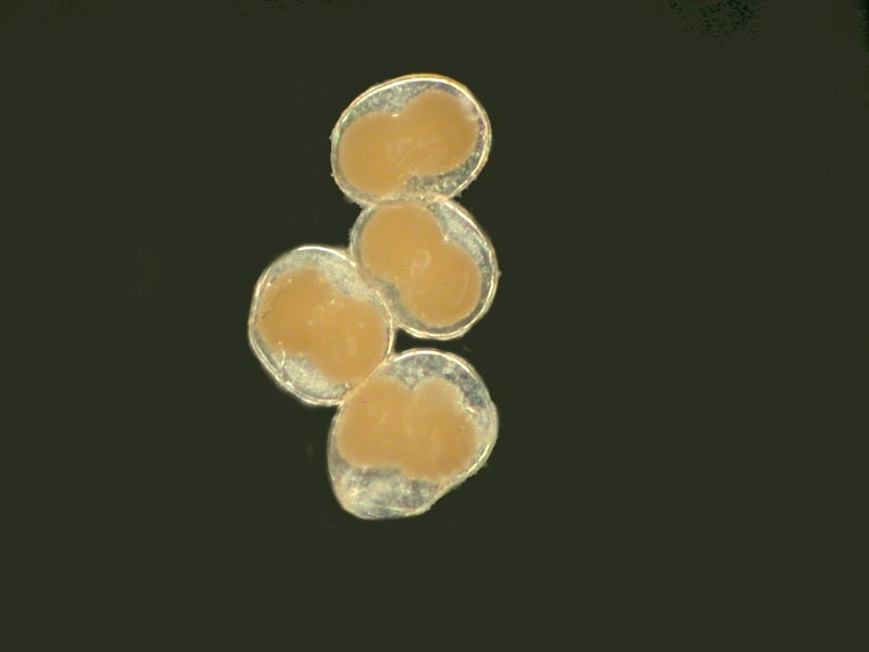
Earlier on, I mentioned how eggs frequently can be found and once again to emphasize the delight of toying with the lighting, I will show you 3 examples of one egg cluster and I have no idea what they might be.
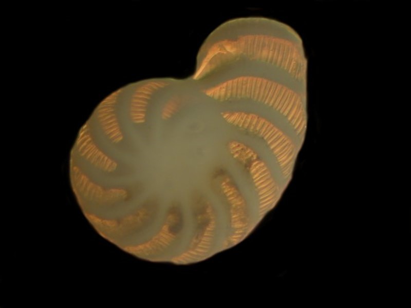
And finally, what would a sample gallery be if it didn’t include at least one splendid foram?
This is only a small selection of the marvelous micro and macro wonders that can be found in such collections. So, if you have some preserved samples sitting around, get them out and you will have the possibility of many hours of entertainment.
Who knows, you might even discover something entirely new.
http://www.bbc.com/news/science-environment-29054889
(Thanks to Dave Walker for this reference.)
All comments to the author Richard Howey are welcomed.
Microscopy UK Front
Page
Micscape
Magazine
Article
Library
Published in the November 2014 edition of Micscape Magazine.
Please report any Web problems or offer general comments to the Micscape Editor .
Micscape is the on-line monthly magazine of the Microscopy UK website at Microscopy-UK .
©
Onview.net Ltd, Microscopy-UK, and all contributors 1995
onwards. All rights reserved.
Main site is at
www.microscopy-uk.org.uk .