|
Black and White Photography
By Steve Durr
|
My first attempts at Photomicroscopy were with Black and White materials and were, to say the
least, of dubious quality. I decided to enrol on a basic course in photography which included the chance to do
your own Black and White printing. Enrolling at one of the local colleges enabled me to gain access to a dark room
and also some professional help. Building your own dark room is not that difficult but actually running one at
home can become quite expensive. If you persevere and stick at the course you should be able to knock out some
half decent prints within the year.
|
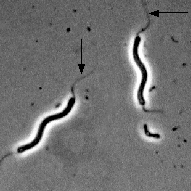
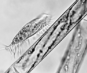
This photograph is of a fresh water microbe and shows the flagella at the two ends of the
cell marked with an arrow. The flagellum helps the organism to move around in the liquid medium. Phase contrast
enabled me to pick up the fine detail which would other wise be lost. X 1000.
|
|
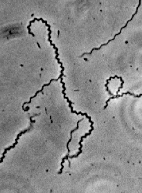
This spirochete was photographed using phase contrast, and was also taken without using flash.
Because I do not have flash, taking a photograph of something that never wants to stop can take an awful long time.
|
|
|
The two films that I use are T-Max 100 and Technical pan rated at 64 Iso. When light levels are
really low then T Max 400 can be used.
Many modern cameras are not much use for taking photographs down the microscope because of the
built in automatic features. A much better way is to try and buy a second hand Olympus OM-1 or OM-2, both of which
are superb for attaching to the microscope. They support many extras including the interchangeable focusing screens,
which are a must when high magnifications are being used. The more expensive option is to buy a microscope which
has a built in camera and more or less takes care of the exposures for you.
In order to obtain the best quality negatives when using B/W emulsion, a good working knowledge
of filters and their use is of paramount importance. Filters can be made of glass or more commonly are sold in
the gelatine form. Care must be taken with the latter type because they can be easily scratched. The use of filters
enables the photographer to be selective about what part of the photograph is going to be enhanced. This prevents
the negative from being very similar shades of grey. The drawback to all this is that filters absorb light and
therefore exposure times increase. This increase in exposure time can be a nuisance when photographing live animals
that move.
|
| This photograph is of the centrally aligned nucleus of the alga Spirogyra, the nucleus is held in place by the cytoplasmic strands which can be seen radiating outwards toward
the cell wall. Reproduction is by fragmentation and also by the formation of spores, which sink to the bottom of
the pond to await more favourable conditions. |
|
|
| |
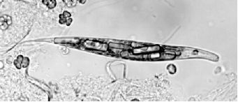
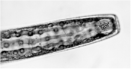
| This species of Euglena seems to
be very common in the ponds of Epping forest. The large rectangular shapes that can be seen embedded within the
cell are for storing carbohydrate and are called Paramylon bodies. The Red eye spot and the flagella are at the
anterior of the cell, these two organelles help the organism to manoeuvre about in the water. |
|
|
|
|
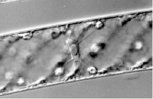
This type of photograph relies on sheer good luck rather than any planning and should be seized
upon immediately. This is a hypotrich ciliate called Euplotes and was photographed wandering down the filament of Spirogyra. The ventral cirri that appear to look like
legs are in fact modified cilia and they help the organism to move over the substratum. |
| This shows part of the Desmid called Pleurotaenium. The crystals, which can be seen in the
two small vacuoles at each end of the cell were, for many years, thought to be of calcium sulphate, but as more
work has been done on the composition of these crystals it has since been learnt that they are in fact made from
barium sulphate. |
|
|
It is always a good idea to use a Yellow Green filter when taking photographs down the microscope
in B/W. When using phase contrast a green filter should also be employed. These filters will help to overcome the
inherent defects of the achromatic lens and should produce a sharper image and therefore better contrast.
Black and White processing can be great fun and can teach you much about how images are formed.
It also allows you to have the final say into how you wish to present your image by the use of filters and the
actual processing of the film. With the advent of image analysis techniques and computers that can be linked up
to cameras this type of wet processing may not be around for much longer.
Why not take a closer look at the differences between protozoa and bacteria and pay a visit to
Wim Van Egmond's The smallest
page on the web.
Comments to the author Steve Durr are welcomed
Please note: this is a free resource provided by Microscopy-UK. We have worked for 7 years without
pay to create one of the most content-rich sites on the web. Our costs are increasing. If you believe this resource
is worth keeping freely available to all, perhaps you might wish to consider donating just a small amount to help?
Please click here if you might
like to consider a small donation.
It would really help!
Prepared for the web by Anne Bruce
© Microscopy UK or their contributors.
Please report any Web problems to the Micscape Editor.
Micscape is the on-line monthly magazine of the Microscopy UK Web
site at http://www.microscopy-uk.net
© Onview.net Ltd, Microscopy-UK, and all contributors 1995 onwards. All rights
reserved. Main site is at www.microscopy-uk.org.uk with full mirror at www.microscopy-uk.net.