One of the delights of freshwater microscopy is finding the unexpected. Here's an example from my recent casual pond dipping.
For an evening's microscopy I'd decided to look at cytoplasmic streaming in Canadian pondweed (Elodea canadensis), which I'd read showed the phenomenom quite well. So I duly took a small piece of the weed from my garden pond, cut a tiny piece of leaf off and made a temporary mount in a drop of pond water with coverslip. I particularly wanted to see if I could capture a video clip of the streaming ... but I spent the evening studying something entirely different.
When examining the leaf cells, I noticed the entire leaf fragment moving, so I searched the slide to see what little critter had also been inadvertently collected. It wasn't a large protozoan browsing around the leaf as expected, or a small water flea, but a sessile rotifer on the leaf edge. It was a delightful specimen which I think was a species in the genus Collotheca. (Update: Readers have kindly sent in their own comments and images which suggest this is the correct genus).
I believe many members of this genus are quite
common but hadn't encountered examples myself before. So the evening's
viewing was spent admiring this rotifer instead, trying various types of
lighting to show the wonderful detail. I've presented a video clip and
stills below, but they don't really capture the fascinating features of
this rotifer. Watching the mouthparts extend is particularly fascinating
when viewed live.
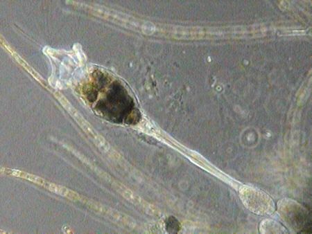
| Sessile (i.e. not planktonic)
rotifer (Collotheca sp.) shown extending. Phase contrast lighting,
10x Cooke phase objective, no eyepiece.
The rotifer extension is in real time (although the repeat extensions aren't as the video is auto-looped). It's quite an obliging 'film star', as a light tap on the stage causes it to contract before it slowly extends again. Thus allowing repeat 'takes' if the video clip didn't work! (485 kbytes, 15 frames/sec). The video is an animated gif (352 kbytes). |
Note: The image may remain
a still until
|
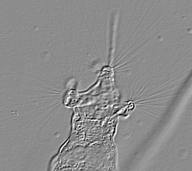
| Larger video still of
above. The setae (stiff hair-like structures) are just visible at the upper
left radiating from the lobes. The three oval objects bottom right may
be resting eggs of this rotifer.
Apparently Collotheca species usually have a gelatinous case, but if present here, it's hard to see for this specimen - even when using darkfield or phase illumination. Fully extended, this specimen was ca. 520µm long (including the mouthpart 'extension' - the out of focus line top left). |
 |
| The very fine setae
were hard to show in an image captured direct from the microscope, even
using phase contrast, so I've cheated here. A brightfield image was taken
with a Russian 20x objective, and the 'emboss' effect used in image software
(Picture Publisher v7.0). This gives an effect which has been dubbed the
'poor man's differential inteference contrast'!
The setae are the fine lines radiating from the lobes. Donner in his wonderful book (ref. 1) effectively describes them as 'the sinister net'. The out of focus vertical line below the central lobe is a thicker feature extending beyond the lobes. |
 |
If collecting from a pond, don't forget to take some small samples of pond leaves, fine roots etc as these often have many examples of various freshwater groups like protozoa, rotifers, diatoms. My tiny garden pond regularly contains green hydra for example, which aren't collected from a plankton net sweep, but which can be found attached to the underside of floating pond leaves.
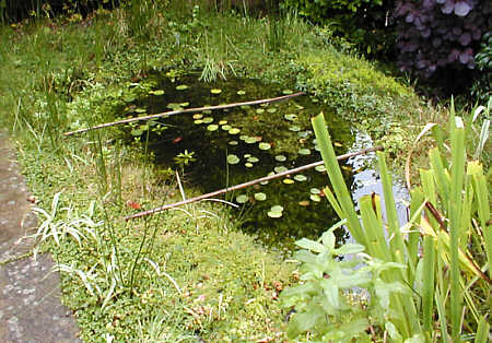
| The 1.5m long pond in
the author's small garden. No ornamental fish are kept in order to encourage
natural aquatic life. But was pleased to find six frogs have taken up residence.
Larger plankton like water fleas don't seem to have become established, but there's plenty of smaller pond life, especially attached to the weeds as described above. |
 |
(Update Oct. 26th. Acknowledgement: Many thanks to Mike Morgan, Steve Durr and Wim van Egmond who have sent me splendid images of Collotheca from samples they have collected.)
Further reading
'Rotifers' by
Josef Donner. Translated and adapted by H G S Wright. Warne, London,1966.
Now out of print,
but this is a splendid introduction to the rotifers. The many line drawings
often allow a provisional identification of a rotifer to family or genus.
'Fresh-water
Invertebrates of the United States' by R W Pennak. Third edition, Wiley,
New York, 1989. Chapter 6 covers the rotifers, with many illustrations
and a key to genera.