
by Rosemarie Arbur, Oregon, US |
|
by Rosemarie Arbur, Oregon, US |
| Actinobolina is a
ciliate of medium size, 100-200 µm long. It's oval, mostly transparent,
vacuolated, with faintly visible longitudinal rows of body cilia. A first
and even second glance do not necessarily reveal its identifying characteristics:
the sausage-shaped macronucleus that sometimes looks like a mere opacity
among the vacuoles, and the "tentacles" that look like its regular cilia,
only three times as long. Three months of finding little but small-to-medium-sized,
oval, mostly transparent, vacuolated protists with sometimes-visible rows
of cilia kept me on close terms with the drawings in my reference books.
Poring over drawings caused
"Actinobolina vorax" to be one of the phrases I heard inside my
head whenever I found yet another oval ciliate. In June, I had drawn a
rough oval with a very plain cytostome at one end and a water-expelling
vesicle at the other; it was supposed to represent a 200-µm-long
organism that had a rotifer inside it and a 140-µm-long one that
had paused long enough for me to discern the gymnostome mouth. Two months
later, when I found the critter below, my subvocalized "Actinobolina"
mantra reminded me to look at it very carefully.
|
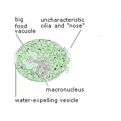 |
When I first saw this organism, it was 200 µm long. It had a circlet of cilia very near its front end and what looked like tiny cilia on its protruding "nose." (Trouble is, Actinobolina are not supposed to have those kinds of cilia at those locations.) But it was the right size, it had one water-expelling vesicle in the right place, it had a large food vacuole inside, and it even had what looked like an Actinobolina's macronucleus. Before I could confirm and add to these observations, the critter began to do weird things. |
 |
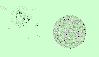
It lengthened itself and created an indentation around its middle. Its front end squared off and flattened, as if it were pushing itself hard against the slide or cover glass. The lengthening and indentation suggested fission, but |
| instead of becoming daughter cells after an unhurried process, the Actinobolina burst apart, ejecting the large food vacuole along with quite a bit of other cytoplasm. Then, as the membrane around the food vacuole broke and the vacuole's contents dissipated, | 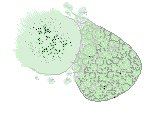 |
 |
what remained of the Actinobolina began to repair itself. Within two minutes, the right half of the original organism became a self-contained spherical body, full of tiny vacuoles that obscured whatever was inside. |
| Five minutes more brought visible cilia, rotation around an axis one end of which seemed to be the mouth, larger and more transparent vacuoles, a defined macronucleus, a change from sphere to ovoid, and a length of 160 µm. | 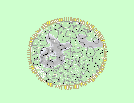 |
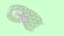
| The Actinobolina continued to refine itself. Changing my illumination from dark field to bright field to oblique and back again, I think I got to see most of its visible features. The upper half of this drawing omits most of the vacuoles to show the rows of cilia, and the lower half omits ciliary rows to emphasize the many small vacuoles among the larger, more transparent ones. |
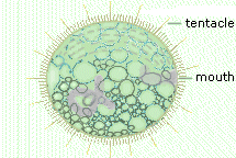 |
| Looked at straight on, the mouth is a simple oval; from the side it appears elongated and denser than the cytoplasm around it because it's a clathrum, a reinforced orifice common to the class Gymnostomea that can expand greatly to accommodate large prey. Its location at the front of the body and the way the rows of cilia converge toward it help distinguish Actinobolina visually from the larger also-oval members of the family Tetrahymenidae. Just as distinctive are Actinobolina's tentacles. They look like long cilia but are functionally quite different: they contain toxicysts which, when they touch Actinobolina's prey, paralyze it and thus make it easier to ingest. According to the reference books I consulted (Jahn, Kudo, Pennak, and Rainis), Actinobolina's normal shape is a longer oval than I show here. This one, after all, was still recovering from the loss of 25% of its former self. |
| Afterthought Readying this article involved quite a bit of re-drawing. On a hunch, I re-colored the anomalous "nose" of the first illustration, andlookI bet my Actinobolina had been eating another ciliate, had been being truly "vorax"! ... Was its prey more than it could hold? | 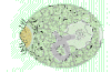 |
Comments to the author Rosemarie Arbur welcome.
Please report any Web problems or
offer general comments to the
Micscape
Editor,
via the contact on current Micscape
Index.
Micscape is the on-line monthly magazine
of the Microscopy UK web
site at Microscopy-UK
WIDTH=1