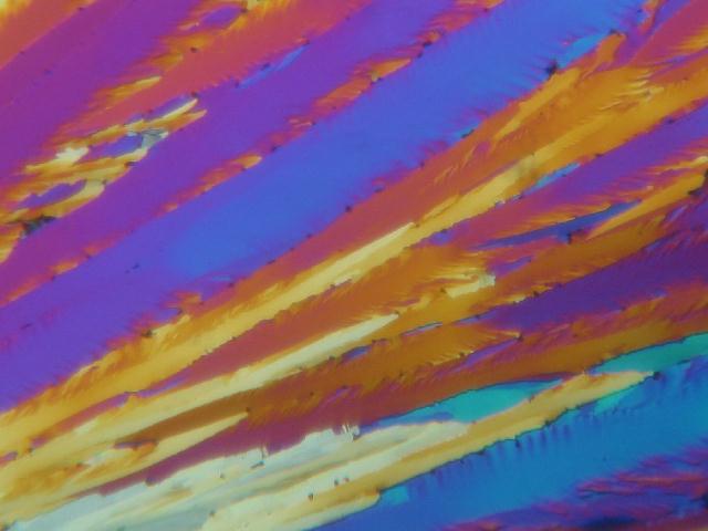
Many objects that are dull and uninteresting, can be enhanced by being viewed in polar illumination. Sometimes parts of an organism can be revealed that were otherwise almost invisible. Two polar filters are required, one in the filter tray below the stage and another in a convenient place in or below the eyepiece. Before objects are placed on the stage, one of the filters must be rotated until no through light is visible, when looking down the eyepiece. A fairly strong light source is required. The insertion of a selenite slip or even pieces of plastic over the substage filter can dramatically alter the picture.COUMARIN.
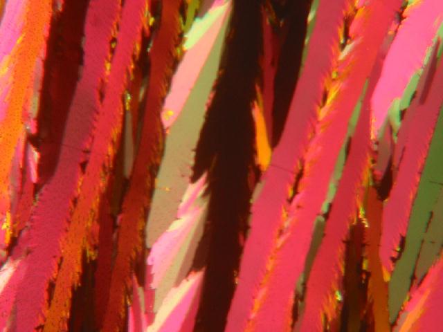
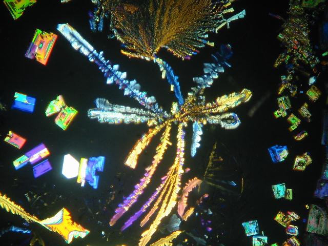
This is a less common substance, to be found mounted on old microscope slides. It was sold as either a "Rainbow" of colours in a curved multicoloured display or as can be seen below as dendrites in many hues. There is little doubt that coumarin is one of the most colourful objects for the Polariscope. The actual substance occurs naturally in the sweet-clover and can be smelled in the aroma of newly mowed hay, but it has a bitter taste. The technology for the production of these crystals is not generally known by the amateur microscopist.
THE UPPER EPIDERMIS OF DEUTZIA SCABRA.
This was a very popular object stripped from the upper epidermis of the leaves, and mounted in Canada Balsam. When viewed in plain light there is little of the leaf hairs to be seen, but under crossed polars they sparkle quite brilliantly. In this slide a blue selenite slip has been used to impart a background effect.
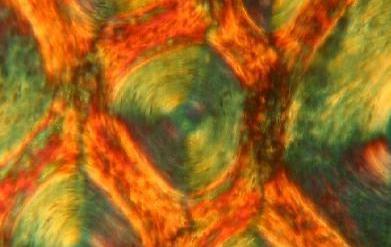
SECTION OF RHINOCEROS HORN.
This is a very small horizontal section of the rhino horn. The horn is much prized in Africa, and is not generally available. Polarisation reveals that the horn is really a hardened gathering of hairs. The detail is well displayed by turning on a rotary stage and selenite filters add to the display.
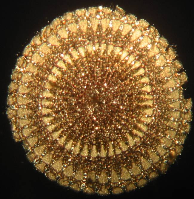
SECTION OF AN URCHIN SPINE.
These sections were an essential component of the Edwardian collection. These were sawn off from large spines into thin discs, which were then finely ground down to very thin sections. Not all urchin spines are suitable for this work.
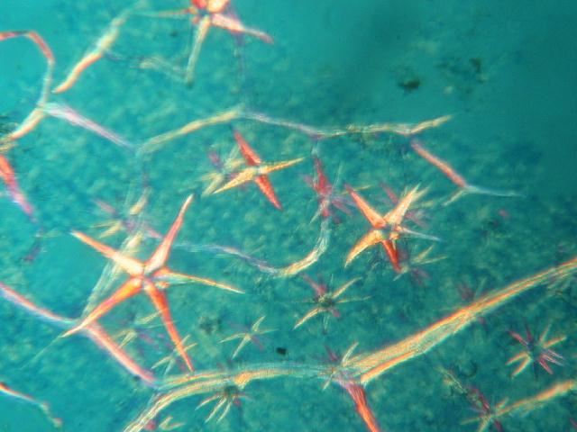
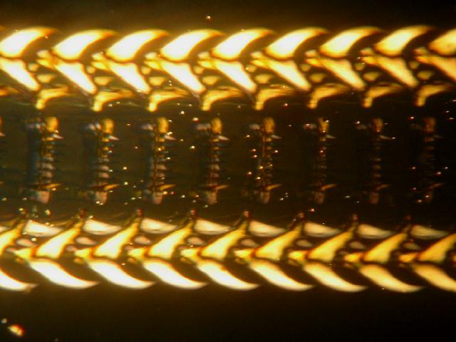
STRONTIUM PLATINOCYANIDE CRYSTALS.
These crystals are particularly beautiful and numbers of different shapes can be found on one slide. The raw material for this work is now not freely available to microscopists in the UK, but it has to be one of the most diverse for shape.
THE RADULA OF A WHELK.
This rasping organ which is used rather like a flexible file is used to graze seaweed. For a mouthpart it constitutes a formidable tool. Viewing is greatly improved by the "blue" selenite slip. The whelk radula can be mounted after extraction and cleaning from the "mouth area" by dissection and immersion in a solution of sodium hyroxide, although this substance is very caustic, and very dangerous to the eyes in particular. It should only be done by those with experience in handling caustic materials in a safe place like a fume cupboard.
Comments to the author, Brian Darnton, are welcomed.