When one has a passion for natural history, a curiosity about exotic and unusual organisms, and only modest financial means, one must be inventive and hunt for hidden treasures in creative ways. At a yard sale, at the age of 15, I bought a quart jar of old shells for 25 cents, probably remnants from some long ago vacation trips. To the casual eye, they looked quite ordinary, but I had glimpsed a few broken shells, two broken sand dollars, and several sea urchin tests (shells) which looked like they had been caressed with a hammer, in short, they were in fragments. I was delighted.
The lantern (jaw apparatus) of a sand dollar.Now, you may think I am in need of some extensive psychological counseling, but have you ever thought about what the inside of a shell looks like with all its wonderfully smooth and glistening chambers? When I got home and spread out my loot to examine it, I was at once intrigued and frustrated. The broken shells were sheared at odd angles and didn't satisfy my sense of symmetry which a nice clean cut down the length of the shell would have provided. At another yard sale, I had purchased, for the grand sum of 2 dollars, a jeweler's saw with 6 blades. Four blades, three blisters, and five hours later, I had two very nice halves of a 2 inch shell. Only years later did I discover that one could purchase beautifully cut shells for a modest price. Nonetheless, the experience was, for me, enormously satisfying and, as I gazed at the whorls that spiraled outward from the tip of the shell, I felt a deep pleasure at observing the internal symmetry and, as a consequence, symmetry has been a source of fascination for me ever since. Anyone who shares this enthusiasm, should have a copy of D'Arcy Thompson's On Growth and Form published in the early part of the 20th Century. Just a few years ago, Dover Press reissued, in a paperback edition, the entire 1000+ pages for only about $25. Hurrah for Dover! Also, shame on Dover. The one drawback is that it does not have the excellent binding that characterizes most of their other books and it will crack and start to come apart, but then, I suppose, these days that had they done the crack-proof binding, it would have cost $125 instead of $25. A very good compromise is the abridged edition of On Growth and Form done by the distinguished developmental biologist, John Tyler Bonner. Professor Bonner's careful editing produces a very readable text and omits errors, many of which are simply a consequence of new research and knowledge acquired since D'Arcy Thompson's time. If you can afford it, buy both versions. The Bonner version gives you the essence, the full version has all the warts and wrinkles that show an extraordinary man wrestling with the whole character and future of biology in the 20th Century. This book is one of those classics like Darwin's On the Origin of Species, often referred to and rarely read.
But back to my jar of shells and the two broken sand dollars. Sand dollars are like sea urchins all sort of flattened out and they tend to burrow in the sand, thus their descriptive name. Sand dollars are covered with thousands and thousands of tiny spines. If you want to know exactly how many, take one and put it in a jar containing a mixture of liquid household bleach and water and all the spines will fall off and you can count them. Be careful, the bleach is caustic and you should never mix it with any other chemicals or household products unless you know precisely what you are doing. Certain combinations can produce lethal gases. However, bleach diluted with water is safe enough as long as you dont try to drink it, use it as eyewash, or bathe in it. The wonderful thing is that you not only get all those spines to count, but you get a nice white, bleached sand dollar shell which allows you to examine the intricate structural designs underneath all those spines. If you look at several genera of sand dollars, you will notice significant variations in shape, size, thickness, whether or not there are lunules (elongated holed in the shell), and the patterns of the petaloids which are flower-like arrangements of holes through which the tube feet extend. Tube feet? Yes, one of the remarkable things about the echinoderms, all those spiny-skinned critters, is that they possess tube feet as a part of a water vascular system, which is an engineering marvel, but thats another whole story and we need some living (or, at least, whole, preserved and unbleached) echinoderms to investigate that topic.
So, back to the broken sand dollars. If you take a piece and look carefully inside, you will see that it looks like a parking garage or even better a section of the bone of a bird wing. There are all kinds of struts and pillars running from floor to ceiling providing strength, durability, yet lightness and space; for, after all, the sand dollar does have to live in there. If you have a species with a thin shell, then you can use forceps or very small needle-nose pliers or the plier-like toenail cutters and slowly chip and trim away pieces until you can gradually get an idea of the floor plan of the sand dollars residence.
If you have a thicker sort of species such as an arrowhead sand dollar or sea biscuit, well, they you can dig out your trusty jewelers saw and go to work cutting both cross sections and transverse sections. It is best to do this away from your microscopes as the process tends to produce a lot of very fine white powder that will settle on lenses, the inner workings of mechanical stages and other such delicate parts. If you take some of this powder, dissolve it in a weak solution of a drop of acid on a slide and let it dry, youll get some splendid crystals, which when place under polarized light, will give you a miniature light show in color. You can also try this with some of the spines, after youve counted them, of course.
As you explore the surface of sand dollars, you will notice tiny bumps and pits where the pines were attached. The thick end of the spine and the surface bump are essentially a ball and socket joint. This can be more readily observed in a sea urchin where the bumps are more prominent and the socket at the base of the spine is more evident.
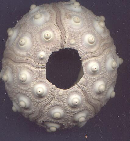
The surface of an urchin with spines removed.Echinoids (which term is derived from the Greek word for hedgehog) include sea urchins, sand dollars, sea biscuits, sea cookies, and heart urchins among others. When I pulled a piece of a broken urchin out of the jar, I was amazed to discover that the inside was very smooth, but perforated by tiny holes through which the tube feet extend. On the top surface, the rows of balls for the ball and socket joints showed up very distinctly. If you now begin to examine the test closely, you will open up a whole new world and discover the periproct, the anterior ambulacrum, the posterior interambulacrum, perhaps even some remains of triphyllous pedicellariae.AAAARGH! Yes, the terminology associated with echinoderms is thorny indeed, but, in part, this is due to the fact that there are so many intricate, fascinating, and sometimes minute structures. Find a good introductory textbook on invertebrate zoology and plunge. You will discover that some of the terms like lunule and petaloid are descriptive and thus quite helpful and easy to relate to. Other terms sound like those that a machinist might use, for example, a typical sea urchin spine consists of a shaft, a collar, and a milled ring. With a bit of patience and persistence, you can soon master the basics and its well worth it.
Let me just mention a few of the structures that have particularly caught my attention. Lets start with one thats absolutely basic to sea urchinsspines, which come in an enormous variety of shapes and sizes. (If one argues for the existence of God on the basis of design, then when it comes to sea urchins, it would be difficult not to conclude that God is a slightly mad exterior decorator given to a weird combination of Rococo and Art Deco.) Spines can vary in size from less than an inch to more that a foot in length. Many are sharply pointed and certainly dont seem like an advertisement for a tasty meal. Consider, for example Arbacia, the common toothpick urchin. You might pluck a spine to clean between your teeth after a meal, but you would certainly have to very hungry indeed to chomp down on one of these sea porcupines. Ah, but inside, there are soft, sumptuous treats to be had, as some sea birds have learned. They fly down and pluck up an urchin, soar above a flat of rocks, and drop it, repeatedly if necessary, until it cracks open and all the tasty parts are vulnerable and available for a quick meal. Fast food, ocean-stylebut I somehow doubt that the McUrchin sandwich will ever quite catch on.
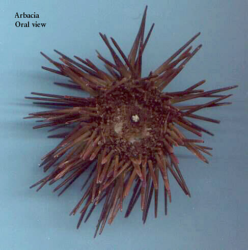
Arbacia (the "toothpick" urchin).The spines of some urchins are poisonous and emit a toxin when the epithelium of the spines is touched. This toxin is generated by a sac-like structure and the poison is reported to be very painful. There are also certain types of spines, such as those of Strongylocentrotus franciscanus (isnt that a lovely name?) which consist of rings of small flat crystals, rather like a row of elongated flat fingernails encircling the central column and there is row after row as you move up the axis of the spine. The most interesting thing is, that if you take one of the spines, which can be several inches long, and run your index finger and thumb from the base to the tip, your thumb and finger glide along the shaft with no difficulty. However, when you try to reverse this movement, you have a very different experience; it feels like you are trying to move your thumb and finger down a spike covered with minute thorns projecting upward. Imagine stepping on one of these urchins!!! Such a device certainly will discourage the majority of would-be predators. These spines are fascinating to look at with a stereo microscope, as the crystalline structure is readily evident and they have a rich glossy, burgundy color.
Before I forget, let me mention that the cross sections of echinoid spines can be among the most beautiful of microscopic specimens. If, however, you attempt to produce a section yourself, you will very likely regard the price of commercial slides as a real bargain. First, you need to find a nice, well-formed spine; then you need to find a fine-toothed saw blade that will fit in the handle of a craft knife (perhaps, the trusty jewelers saw will work, if the spine is sturdy enough). The next step is to cut a section with the saw that is as thin and flat as possible. This is not a simple task; some spines will shatter, S. franciscanus for example, while others will break jaggedly just as you are nearly finished cutting the section. If you get lucky and have a cooperative spine and a saw that is giving you good results, then cut a series of sections, because the chances are good that youll need them. Next, take a knife-sharpening hone with a medium grit and begin the process of grinding the surface flat. Some mounters used to glue the section to a slide with balsam and then grind, dissolve the balsam, glue the other side of the section to the slide and grind again. This whole process is repeated using a fine grit stone and finallyif your section is still intact, the finishing touch of polishing is done with extra fine abrasives between two slides.
I use a somewhat shorter method and the results are sometimes a bit uneven. (Maybe when I retire, Ill find time to do it in the exacting manner that produces nearly perfect specimens and then Ill sell them all to you for $75 a piece.) Even my shorter method is, however, rather tedious, so dont expect miracles. Once I get a good section cut, I grind it on a small fine diamond hone. You can buy these at a discount store for $5 or $6. If the spine section is very small in diameter, you will probably have to glue it to a slide with balsam or an acetone-based household cement. This does have the advantage of allowing easier manipulation of the section and also strengthening it, since the balsam or cement penetrates the empty areas of the spine. The disadvantage is that it can make the section sticky and powdery as you grind. The spine is made up largely of calcium carbonate (chalk) and as you grind, a fine dust is produces that easily adheres to the cement and then makes further grinding to produce a nice flat section, more difficult.
I much prefer to work with types of spines that have a larger cross section; with these you simply take your right index finger and move the section over the hone. As the section gets thinner, ease up on the pressure in order not to break the spine. You do, of course, want to grind both sides. This method also has the advantage of removing the fingerprint from your right index finger, which is very handy when you commit a major felony burglary, stealing rubies, diamonds, emeralds, etc. from the rich. (Dont take any microscopes they might havethat would be a sin!) You do, of course, have to remember while committing this felony, not to touch anything with anything other than your right index finger. Actually, a small adhesive band-aid can reduce wear and tear on that precious finger.
As the section approaches appropriate thinness and flatness, place it in a small dish and direct a stream of water at it either from a wash bottle or a series of streams from a pipet. This helps wash away the chalky material which has accumulated and allows you to make an overly optimistic estimate of how many hours of work you have yet to do.
From here on in, things are very much touch and go. The next step for me is a fine emery board, such as one might use on ones fingernails. This can be quite handy in smoothing out irregularities on the surface, but one must be very careful, for a little too much pressure and the section will break. (I speak from frustrating, angry, furious, depressing, enervating, maddening experience.) Sheets of very fine emery paper can also be purchased and are quite useful. In addition, there is a very fine abrasive called crocus cloth which can also be used in the near final stages. Another item I have found helpful is special cytological slides, one entire side of which has been ground. The section can be placed between these and gently ground with or without powdered abrasives. Finally, the very finest abrasive powder you can obtain can be used between two plain glass slides and then ,the final polishing is done with just the slides and no abrasive. You can see why such slides are now rarely made and why older ones, when found, are quite expensive.
Some of the early work (cutting the sections) and sometimes even some of the work in the middle stages can be done with an electric hand tool if the spine is sturdy enough. It is best to have a tool with variable speed as this increases your control. The type of spine to practice on is probably one from the so-called club-spined sea urchins. These can be 2 to 3 inches in length and a bit greater in diameter than an ordinary wooden pencil. The initial sections are relative easy to cut and will not shatter as do the more crystalline spines. The rest is a matter of patience, patience, patience and a fair amount of luck. And again, you might ask yourselfwhy go to all the bother? Well, if you ever see a really fine preparation under polarized light, youll understand why.
I started to tell you, before you got me off on the tangent about preparing slides, about the diversity of spines. The ones from small sand dollars can easily be washed, dried, and then used to make very nice strew preparations. We have already mentioned the club spines and there are some genera that have spines which produce a triangular cross section. Many years ago, I purchased from Turtox Biological supply Co. (now unfortunately extinct), two specimens of a tropical urchins of the genus Podophora (Colobocentrotus). This is an urchin adapted for living on a coastline with a heavy, battering surf and its spines are radically modified. Looking down from above, the spines on the top surface appear essentially flat and, though some are four-sided, most are pentagonal or hexagonal. In general, they are less than an inch long and the sides slope down from the top to narrow at the point of attachment to the shell. The ambital spines, that is, those running around that imaginary circumference that divides the top of the shell from the bottom, are also flat and shaped like elongated fingernails.
There are other genera that have spines that look like paddles or spatulas with wide flat ends that again narrow toward the shell. Yet other forms produce bristly spines that have a disk or flattened mushroom-like plates at the terminus. It is difficult to observe the enormous variety for oneself, since biological supply houses tend to stock only about a half dozen kinds of echinoids.
However, this need not discourage you, since within the readily available species, there are some other amazing structure which we havent even mentioned yet. Sea urchins are not what you would call speedy and often, if they find a good feeding spot, they will cling there with their tube feet for some considerable time. Detritus falls on them and could begin to accumulate on the test; larvae of many kinds of organism seek out any spot that seem even temporarily stationary. To counteract these nuisances, many genera of echinoids have developed pedicellariae, which are ingenious and bizarre, tiny pincer- or jaw-like structures on a stalk containing a central rod. There are four basic types and each has a number of variants. The top part or head is usually composed of 3 jaws, but there can be 2, 4, or 5 in some genera. Although, the details of structure are quite different, the general appearance and function are similar to the avicularia found on some bryozoa.
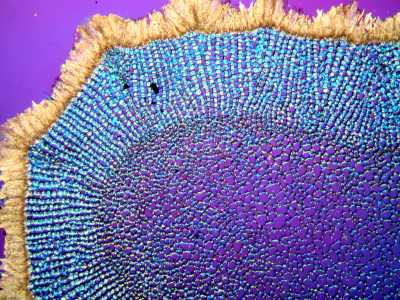
Cross-section of a sea urchin spine under polarized light.The strangest and most intriguing of the pedicellariae are the ones designated as globiferous. Each jaw has a tooth at the tipin some species, several teethand each tooth has a glandular sac connected to the tooth by a duct which can discharge a toxin into any hapless, tiny creatures that intrudes into its arena. Imaginetiny stalked jaws with poisonous teeth hidden down among the spines!
Another odd feature of most echinoid text are sphaeridia. These are to be found in the ambulacral areas and there may be only a few or they may be quite numerous. These tiny hemispheres can be vitreous in appearance or rather dulled by the growth of tissue over them and most have a minute stalk, around the base of which runs a minute nerve ring. In fact, a fine network of nerves runs up along the spine to the tip in most echinoids. The function of sphaeridia is uncertain, but one view is that they play a role in the equilibrium of the urchin.
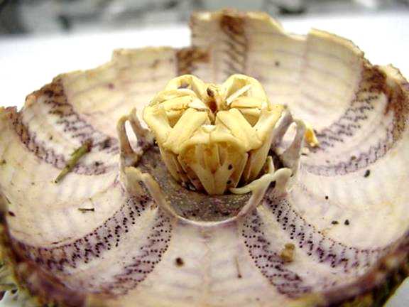
Historically, especially for a philosopher, perhaps the most fascinating feature of all in sea urchins , is the famous Aristotles lantern. Sometimes if you come across an old test, in which you will find bits of the lantern still rattling around inside. In this case, however, what you need is a whole, well-preserved urchin. Turn it upside down and look directly at the center. You will see five little calcareous teeth projecting out. If you break open the test carefully, you will discover a wonderfully complex device for grinding food and this structure consists of an elaborate musculature and 40 calcareous plates! and, yes, Aristotle was actually the first to describe it. He wrote extensively on nearly every subject imaginable, but seemed to have a special passion for animals and wrote widely, and, at times, wildly about all manner of creatures. The Historium Animalium consists of 12 books which survived and it is reported that in total he wrote over 50 works on animals, so the majority of this work is lost to us. Some scientists have argued that Aristotles work retarded the development of science for centuries, but such a claim is simplistic. True, he was sometimes uncritical in his acceptance of reports of bizarre phenomena, such as wind eggs. However, some of his descriptions of marine fauna are so good that we can still recognize the genera today; so he is certainly deserving of having this magnificent little lantern named after him.
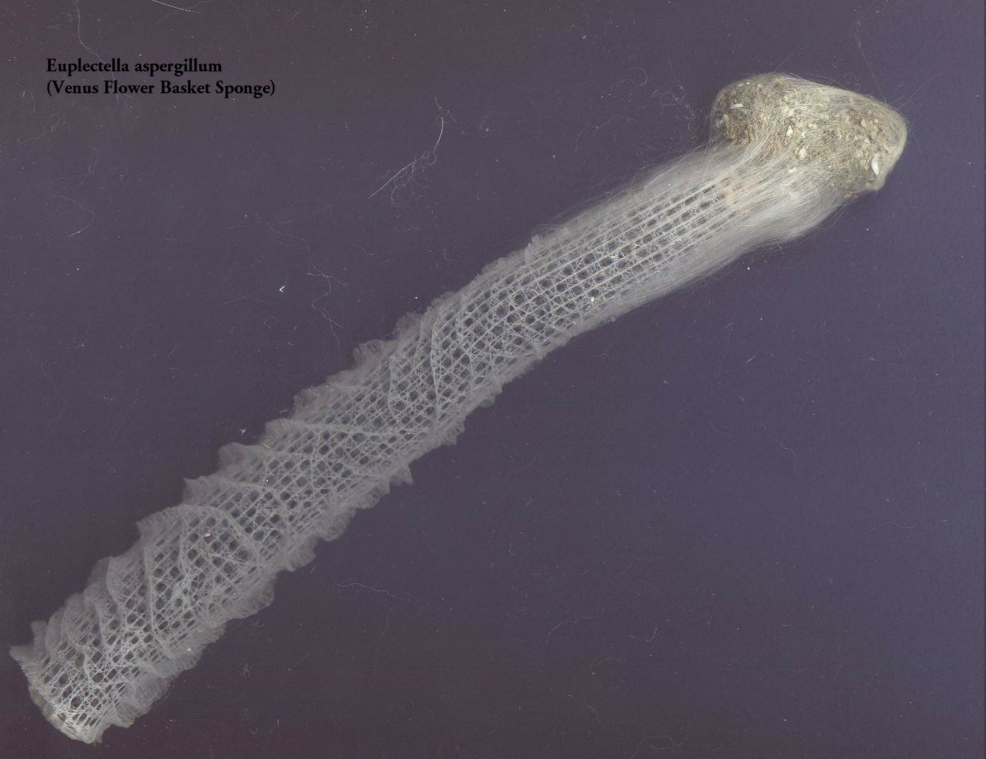
Aristotle's lantern from a sea urchin.I have talked about broken specimens, but I havent yet mentioned dirty specimens. I will mention just one example. Recently I acquired, from a supplier of dried marine specimens, the skeleton of a sponge, Euplectella. This is perhaps, for me, the most elegant biological specimen I have ever acquired. It is also know as Venus flower basket. The specimen is about 8 inches long and 1 and ½ inches in diameter at the top and from there it tapers down almost to a point at the bottom tip. It has intricate lattice work from the top down to the tip where the lattice loses definition and there is dirt and debris and some very long single threads that look like spun glass. As a matter of fact, they are glass and so is the entire skeleton. Yet another description of the remains of these organisms is glass sponge. The dirt and debris and the long threads are a consequence of that fact that this part of the sponge is anchored in the substrate of the sea bottom. Holding the sponge carefully so as not to damage it, use a knife with a small blade or a micro-scalpel and carefully scrape the dirt and debris from the outside of the sponge tip into a Petri dish. When you examine it, you will find bits of the glass threads, clusters of spicules from the sponge, forams, ostracod shells, diatoms, sand dollar spines, and other assorted micro-wonders. if you get lucky, you might get a sponge which has a couple of brown shriveled things trapped inside it. These are a pair of small shrimp which enter the sponge while they are still tiny larvae. As they grow, they get too large to escape back out through the glass lattice surrounding them, but then, why should they want to? Inside this most beautiful of prisons, they have protection and a constant supply of food from the currents of plankton that the sponge cells cycle through. Whey they mate, the tiny larvae they produce are small enough to escape through the lattice and go seek out other glass sponges in which to take up residence.

Euplectella (Venus Flower Basket sponge) with detritus.
(Click image to view a larger version).Much has been written about the complex web of nature and the intricate interdependence of organisms within eco-systems, but this is very easy to forget when you are investigating a particular type of specimen. Always look for attached dirt, debris, gunk, fuzz, fibers, threads, etc.there may be something of surprising interest. When I order preserved specimens from a biological supply house and find them all neat and clean, I am tempted to write them a letter sayingsend me the debris! I doubt that this ploy would work however. I suspect that they themselves scrape off all these micro-treasures, mount them, and sell them to us on slides.
All comments to the author Richard Howey are welcomed.