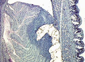
On the back, the so-called mantle, just beneath the
superficial cover, we see a lot of glands in figure 1 which are
dark
blue and greenish in colour.
In fig. 2 we see a flattened space parallel to the back of the
slug. This space is the area into which the glands secrete
their granular products.
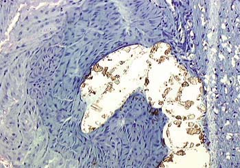
In fig. 3 the cystal-like structure of the secretion becomes
clear.
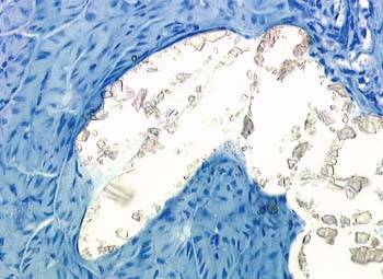
In fig. 4. polarized light was applied to discover whether
the granules were birefringent. They, these crystals,
are accumulated in that flat sac lying in the dorsum of the
slug. Such crystalloid secretions makes a strengthened dorsum,
like a plaque or mantle, to the back of the animal.
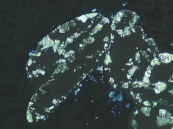
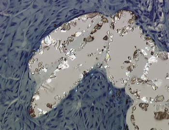
Fig. 5 left. This image was taken between crossed polarisation filters. The shell of a snail (i.e. a mollusc with a spiral shell) may possibly develop in this way by continuous secretion of such crystalloid, calcium-rich material whereas the secretion of it rapidly ceases in the naked slug (a mollusc without a shell).