|
|
A Close-up View of "Common" and (Ambrosia artemisiifolia & Ambrosia trifida) by Brian Johnston (Canada) |
|
|
A Close-up View of "Common" and (Ambrosia artemisiifolia & Ambrosia trifida) by Brian Johnston (Canada) |
The ragweed plant is the source of much misery for "hay fever" sufferers during the months of August and September. This prolific generator of pollen however, goes unrecognized by many. Unlike other plants of the aster family, ragweed has extremely small flowers and the flowers point down, making them almost impossible to see!
Although the structure of both ragweed types is similar, there are differences. Common ragweed in my area grows up to 0.5 metres in height, whereas the giant variety may grow up to 2 metres high. The male flowers of common ragweed (seen in the image above) are joined to the stem by short stalks whereas the giant variety stalks are much longer. Finally, the leaves are completely different, and it is this characteristic that makes identification of the two types an easy task.
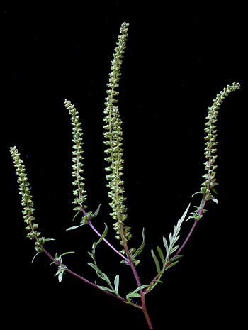
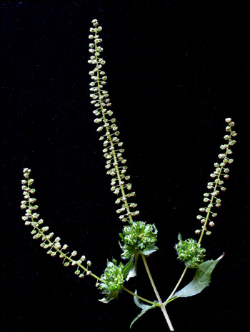
The two images below, of common (left), and great ragweed (right) are not to scale. The flower spikes (racemes) on the left are about ten centimetres in length and the ones on the right are about twenty centimetres long.
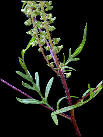
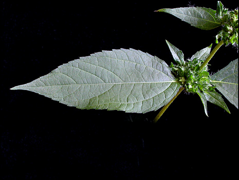
A close-up of the leaves of common ragweed shows them to be finely divided with very deep grooves. Leaves like these are described as highly "dissected". Notice that the plant stem is red near the base of the raceme and green near the top.
Great ragweed leaves by contrast, are described as "ovate" and slightly toothed.
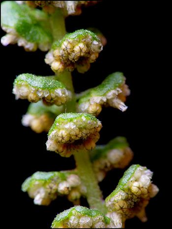
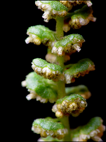
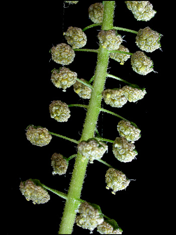
Ragweed is unusual in that it has separate male and female flowers located in different positions on the stem. The male, pollen producing flowers are connected to the spike by a short green stem, and face downwards. As can be seen below, each common ragweed flower-head is composed of many individual flowers, and has a group of green bracts that form a hood over the flowers. The flower-heads are two to three millimetres in diameter. In the image, most of the flowers are unopened, but a few mature blooms display light beige, pollen producing stamens projecting from the underside. The pollen grains produced by these hanging flowers are extremely small and very light, easily caught by the wind and carried over great distances. (A 200 kilometer flight is not unusual!)
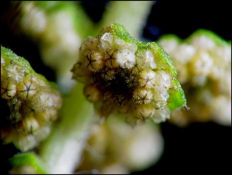
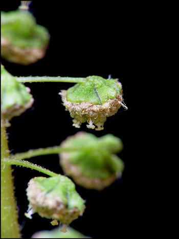
A higher magnification shows that each unopened flower has five petals, clearly visible with dark lines of demarcation between them.
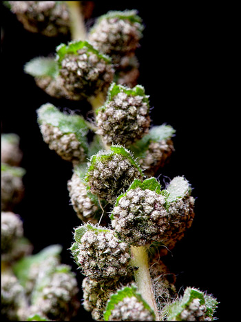
Male flowers at a later stage are shown below. When all of the blooms have matured, the downward facing flowers have a distinctive dark gray colouration.
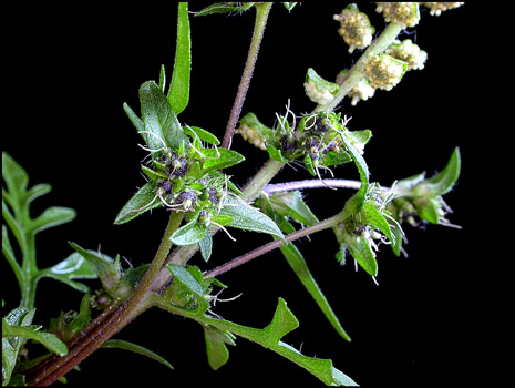
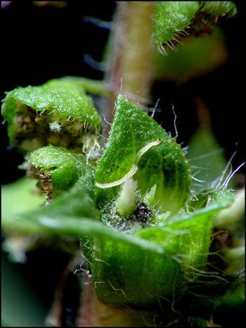
As mentioned earlier, the giant ragweed has its male flowers on much longer stalks. Here again, most of the flowers have yet to bloom, but a few stamens can be seen projecting from the open flowers.
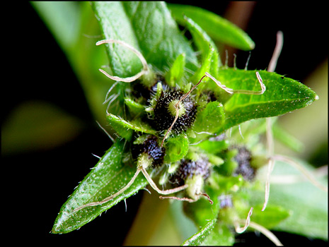
A closer view shows an immature bloom with three open flowers and projecting stamens. The hood shaped bracts appear to cover less of the flower-head than is the case with common ragweed.
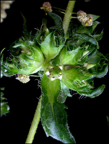
The female (pollen accepting and seed producing) flowers in ragweed plants are found below the top spike of male flowers. They tend to form where leaves meet the main stem. Unlike the male flower-head which is composed of many flowers, the female flower-head contains only one flower. The image below shows a number of these female flowers at the base of a common ragweed spike.
At a higher magnification, three of these female flowers can be seen, each with a distinctive yellow tipped black speckled cone. Projecting out of each cone are the two lobes of the stigma, ready to accept wind blown pollen.
An even closer view reveals one female flower with its green bracts and bi-lobed stigma.
In the great ragweed plant, the female flowers have a slightly different appearance. (The flowers shown are immature.)
Ragweed is a perfect example of an innocuous plant with a "larger than life" impact on the lives of allergy sufferers. Although it doesn't display large colourful blooms, it is interesting by virtue of its strangeness!
Addendum
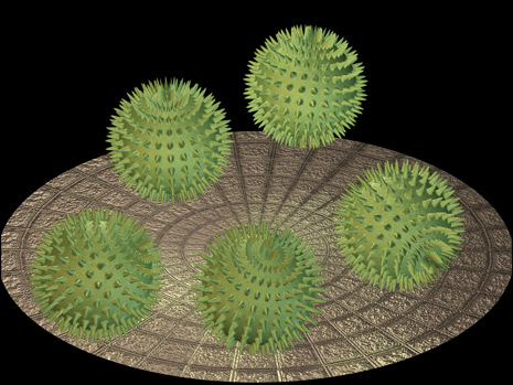
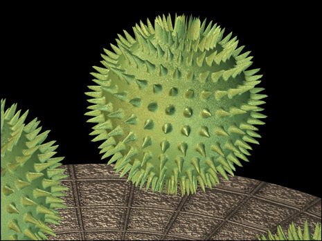
By the time that I decided to write this article using photographs taken during August and September, frost had destroyed all of the ragweed plants in my vicinity. It was therefore not possible to photograph ragweed pollen under the microscope. Instead of sketching a pollen grain, I decided as a challenge to find a mathematical formula that would, when plotted in 3-D, show an approximation of the shape and structure. (A scanning electron photomicrograph of a ragweed pollen grain was used as a model.) One of my hobbies is the study of the three-dimensional representation of complex mathematical functions, and this proved to be an interesting problem. The results are shown in the two images below. For those interested, the equation was first plotted in the software "Mathematica" and the co-ordinate points saved as a file. These points were then loaded into the ray-tracing software "trueSpace 5" and the structure lighted and textured. Note that real ragweed pollen grains would not be perfectly spherical, and the tiny spikes would be more randomly positioned.
The "Mathematica" file to produce a simulated pollen grain is printed below.
Needs["Graphics`ParametricPlot3D`"]
gr = SphericalPlot3D[
2 + Sin[13x]^8 Sin[13y]^8/2, {x, 0, 2\[Pi]}, {y, 0, \[Pi]},
PlotPoints -> {300, 280}, Axes -> False, Boxed -> False,
DisplayFunction -> Identity];
Needs["Utilities`DXF`"]
WriteDXF["c:\\graphics\\PollenGrain.dxf", gr,
PolygonsOnly -> True]
Photographic Equipment
The low magnification photographs in the article were taken using a Nikon Coolpix 4500 with natural light and the Nikon Cool light SL-1. Higher magnification images were taken using a Sony CyberShot DSC-F717 equipped with an accessory achromat which screws into the 58 mm filter threads of the camera lens. (This produces a magnification of from 7X to 9X for a 4x6 inch image.) Still higher magnifications were obtained by using a macro coupler (which has two male threads) to attach a reversed 50 mm focal length f 1.4 Olympus SLR lens to the F717. (The magnification here is about 13X for a 4x6 inch image.)
All comments to the author Brian Johnston are welcomed.
Published in the September 2004
edition of Micscape.
Please report any
Web problems or offer general comments to the
Micscape
Editor.
Micscape is the on-line
monthly magazine of the Microscopy UK web
site at
Microscopy-UK
© Onview.net Ltd, Microscopy-UK, and all contributors 1995 onwards. All rights reserved. Main site is at www.microscopy-uk.org.uk with full mirror at www.microscopy-uk.net .