Some observations and comparisons between two classic ranges.
By Ian Walker. UK.
Introduction.
I recently acquired a set
of four Carl Zeiss Jena apochromatic objectives [designed
for 160mm tube length] which were
incidentally fitted to a Zeiss 'F' stand of the mid 1920's but the
stand
was secondary to my purpose of finding out just how well the LOMO
pre-DIN objectives stand up to the original design made by Zeiss many
years before. In my last article in September 2006 I discussed the
history of LOMO and how the company used the equipment from the post
WWII Zeiss Jena works to build up their own factory and use the
knowledge gained by Zeiss to make their equivalent optics. Due to
problems of mounting my Sony P200 digital camera to the microscope it
would be unfair to take identical pictures with each design and show
them here since slight contrast variations or camera shake could make
one look distinctly better than the other, rather I will make general
remarks from observation with various subject matter but include an
image from the LOMO 20x objective to give an idea of results expected.
General observations.
Although similar there are some
differences between the Zeiss and LOMO objectives. Firstly the Zeiss
objectives are heavily chromed to a very high standard in a very light
gold colour with white paint applied to deeply engraved lettering
whilst the LOMO range are finished and turned by lathe to give a rather
more matte appearance with black paint applied to engraved lettering.
There are differences in the design as well as shown in the following
figures, the 10x NA 0.30 being substantially different whilst the 20x
NA 0.65 and 40x NA 0.95 CC are much closer in features. All the Zeiss
range of apochromatics are engraved with both the focal length and
initial magnification which was typical of the day.
The Zeiss objectives here were made before lens coatings were introduced, with a complex design of maybe ten elements this is bound to reduce contrast to a certain extent especially with the condenser iris at 80-90% of the NA of the objective. Rather surprisingly, considering they are not particularly old, the LOMO optics don't appear to be coated either whilst other parts of their microscopes were using coatings at this time [objectives marked between 1979-1989]. None of these objectives are ideally suited to photomicrography requiring large areas of the field of view in focus due to their limited flatness of field which in the case of Zeiss in the 1920's was not much concern to them in that era when pushing the boundaries of design of objectives requiring high numerical aperture. All the tests were carried out on the refurbished Zeiss 'F' stand with aplanatic oblique condenser set to its centre position throughout.
The Zeiss objectives here were made before lens coatings were introduced, with a complex design of maybe ten elements this is bound to reduce contrast to a certain extent especially with the condenser iris at 80-90% of the NA of the objective. Rather surprisingly, considering they are not particularly old, the LOMO optics don't appear to be coated either whilst other parts of their microscopes were using coatings at this time [objectives marked between 1979-1989]. None of these objectives are ideally suited to photomicrography requiring large areas of the field of view in focus due to their limited flatness of field which in the case of Zeiss in the 1920's was not much concern to them in that era when pushing the boundaries of design of objectives requiring high numerical aperture. All the tests were carried out on the refurbished Zeiss 'F' stand with aplanatic oblique condenser set to its centre position throughout.
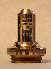
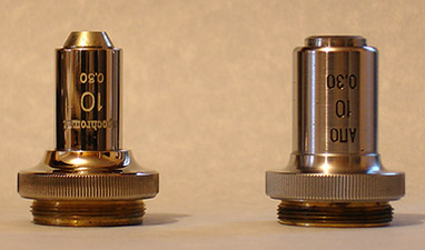
Fig1.
Left and middle, two views of the Zeiss Jena 10x NA 0.30 whilst on the right the equivalent LOMO dated 1980.
Observation with a 10x hand magnifier
shows the first signs of edge delamination in the Zeiss Jena which may
influence contrast. Tests between the Zeiss and LOMO show they are
similar in performance regards perceived sharpness and detail with low
contrast subjects such as diatom test
slides with a tough test of the aplanatic condenser set to 90% of
the aperture of the objectives to show possible problems with flare.
Tests with some well stained Biosil botanical sections show the LOMO is
the clear winner with significantly better contrast, an excellent
performance from the LOMO. Field flatness between the two are roughly
the same. The two objectives are not parfocal probably due to the Zeiss
being physically longer as shown in Fig 1. above. Stopping down
the well corrected three element condenser to 70% of the NA of the
Zeiss objective does not improve
contrast. No signs of
delamination was observed on the LOMO.
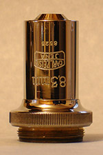
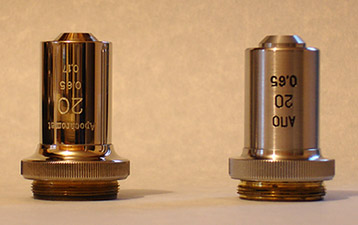
Fig2.
Left and middle, two views of the Zeiss Jena 20x NA 0.65 whilst on the right the equivalent LOMO dated 1979.
Observation with a 10x hand magnifier
shows edge delamination in several planes on the Zeiss Jena which will
definitely
influence contrast. Tests between the Zeiss and LOMO
show they are similar in performance with low contrast subjects such as
diatom test slides. Tests with the Biosil botanical sections show the
LOMO again is the clear winner with much better contrast, an
excellent performance from the LOMO. Field flatness between the two are
roughly the same. The two objectives are not parfocal. Stopping down
the condenser to 70% of the NA of the Zeiss
objective does not improve contrast. No signs of delamination on the
LOMO.
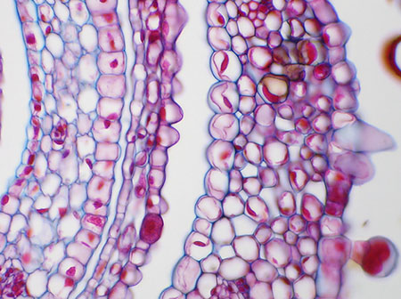
Centre crop from 20x NA 0.65 LOMO objective using a Zeiss 10xK 'Mobimi' eyepiece.
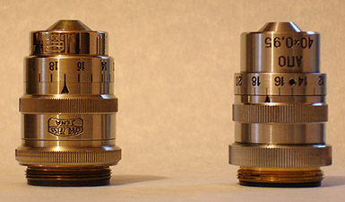
Fig 3.
Zeiss Jena 40x NA 0.95 CC and on the right the equivalent LOMO dated 1989.
The 40x apo Zeiss Jena is finished to an extremely high standard and
unlike the LOMO the collar rotates smooth and free with no backlash.
Observation with a 10x hand magnifier
shows some 'milkiness' to the optics of the Zeiss Jena together with
minor fungal growth between element groups which may
influence contrast. Tests between the Zeiss and LOMO
show they are similar in performance with typical subject matter such
as
a J. D. Moller diatom test slide, in this case experimenting with the
correction collar for best performance which incidentally is much
easier
on the Zeiss because of its free moving collar. There is no definite
winner here, both perform admirably even though the Zeiss optics don't
seem to be in perfect condition. The two objectives are not parfocal
and both need well stopping down on the aplanatic condenser to achieve
good contrast. I could not achieve an acceptable picture on my Sony
P200 due to focus lock being affected by the low contrast of the
subject matter.
Update: Oct. 19th 2006. LOMO stiff 40x apo collar maintenance. Frithjof Sterrenburg in an email discussion, has generously prepared an illustrated guide for the careful worker to correct the stiff collar of the LOMO 40x apo without touching the optics train. This procedure is shown here with Frithjof's permission and with thanks from the present author.
Update: Oct. 19th 2006. LOMO stiff 40x apo collar maintenance. Frithjof Sterrenburg in an email discussion, has generously prepared an illustrated guide for the careful worker to correct the stiff collar of the LOMO 40x apo without touching the optics train. This procedure is shown here with Frithjof's permission and with thanks from the present author.
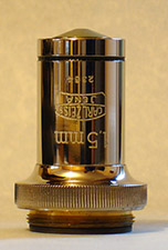
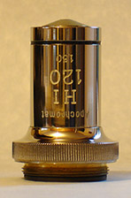
Fig 4.
Zeiss Jena 120x NA 1.30 OI objective, there is no equivalent in the LOMO range.
This objective is described as a 'special objective' in the Zeiss literature for drawing and measuring.
I included a picture of this
objective because it was fitted to the stand and is not often seen
being quite scarce. The more usual oil immersion fitted to one of their
better specified stands would either be the 90x NA 1.30 apo or the
delicate 90x NA 1.40 apo with fragile mounting for the first element.
With some care in lighting technique I can clearly see the line markings on Amphipleura pellucida with this
objective using the Zeiss or LOMO oblique condensers.
Conclusion.
All three LOMO objectives give a very
good performance with no worries of delamination to consider. The Zeiss
Jena objectives are over 80 years old and showing signs of natural
aging; however if your main interest is botanical and histology
sections I would err away from apos altogether having seen excellent
performances from both Watson and Swift
1" and 1/2" achromatic objectives on my Edwardian microscope which are
older still and for the majority of slides give captured images with
flatter field than the Jena's, are easier to use with greater
working distances and work well in conjunction with
the Sony digicam. The best of the three compared would be the 40x NA
0.95 CC
which although not optically in perfect condition still provides
excellent images of diatoms and is much easier to use than the LOMO
with its very stiff correction collar. The 120x OI although described
as a 'special' objective was fairly easy to work with and produced
excellent results on diatom test slides.
A word of caution: Zeiss literature of the time says alcohol should never be used for cleaning the glass on any of the above objectives due to the method of fixing the elements in place.
A word of caution: Zeiss literature of the time says alcohol should never be used for cleaning the glass on any of the above objectives due to the method of fixing the elements in place.