Sigmund Freud’s microscope – on the 150th birthday anniversary of the histologistManuel del Cerro (USA) and Lazaros C. Triarhou (Greece) |
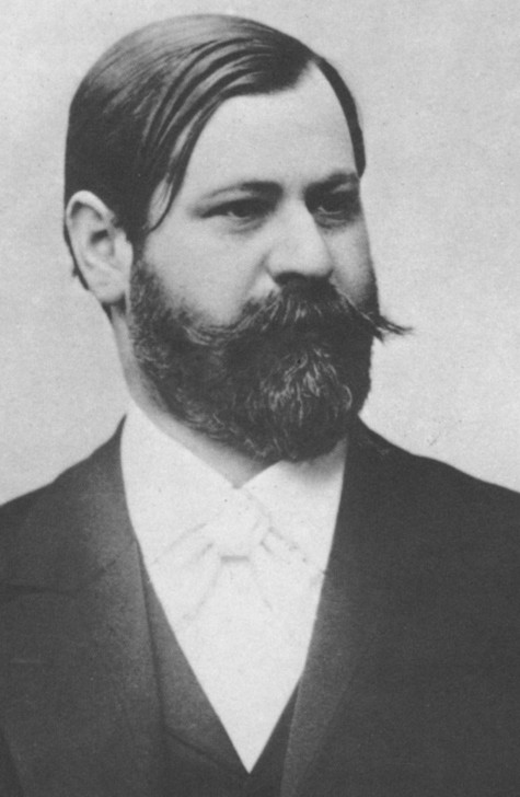
Everyone knows that Sigmund Freud (1856-1939) (Fig. 1) is one of the greatest explorers of the human mind that ever lived (Gay, 1988; Panek, 2004).
Fig. 1. Sigmund Freud in his mid-thirties.
Many people have heard, particularly in Europe, that the year 2006 marks the sesquicentennial (150th) anniversary of Freud’s birth (May 6, 1856); accordingly, 2006 was designated ‘Sigmund Freud Jahr’ in Austria (www.freud-museum.at, www.freud-institut.com) (Fig. 2) and Germany (www.freud2006.de, www.150jahre-freud.de).
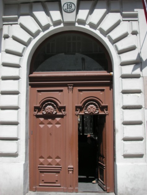
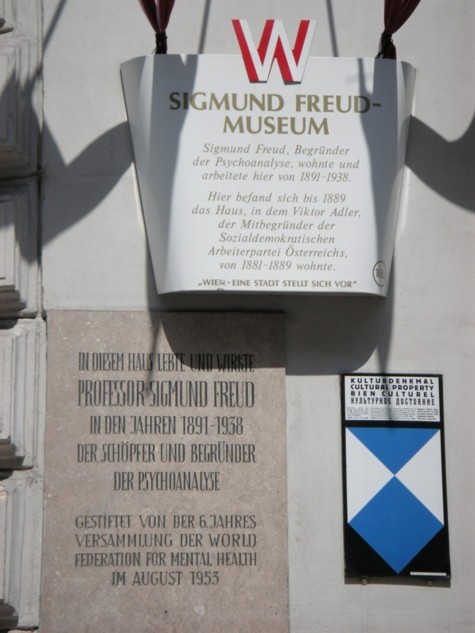
Fig. 2. The Sigmund Freud House and Museum at Berggasse 19 in Vienna.
Photos by one of the authors (LCT).
Much less known is the fact that Freud’s exploration of the mind started with the exploration of nerve cells under the microscope while a medical student. His professional career began in the fields of cell biology and histology (Triarhou and del Cerro, 1985; Triarhou and del Cerro, 1987; Guttmann and Scholz-Strasser, 1998) and gained him wide recognition in neuroanatomy. In this note we shall briefly review Freud’s work as a microscopist, and give details of the microscope type he used for most of his work.
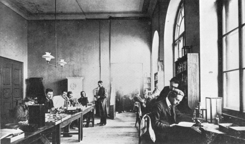
Fig. 3. The laboratory of the Experimental Zoological Station at Trieste,
around the turn of the century. Photo credit Greti Meinx.
From 1875 to 1885, the year he went to Paris to train with the world-famous neurologist Jean-Martin Charcot (1825-1893), Freud worked on histology, neuroanatomy and neuropathology, putting out 14 scientific publications – some constituting pioneering contributions to neurobiology. Nonetheless, the first microscopical study performed by Freud was on a rather unlikely subject: the testis of the eel (Freud, 1877). Nobody however should attach any “Freudian” meaning to this odd choice. The choice was made not by the young researcher, but by his mentor, Professor of Zoology Carl Claus – some things do not change (!) – and carried out by the young medical student during two visits to the Zoological Station in Trieste (Fig. 3).
Original as his first study was, and important as his subsequent studies on cell motility were (Triarhou and del Cerro, 1987), the basic science contributions of Freud that have better survived to our days were those on the structure of the nerve cells (Triarhou and del Cerro, 1985). Those contributions have been highly regarded by established researchers at the time and incorporated into the neuroscientific literature, including citations by Cajal, the father of modern neurobiology, in his classic textbook (Ramón y Cajal, 1897-1904).
Freud conducted his histological studies on the nervous system of the lamprey (Petromyzon) and the river crayfish (Freud, 1878; Freud, 1882) at the Physiological Institute of Professor Ernst Wilhelm von Brücke (1819-1892) at the University of Vienna. He worked in von Brücke’s laboratory between 1876 and 1882. In the legends of his histological drawings in the resulting publications in the Proceedings of the Imperial Academy of Sciences, Freud gives technical information on the optical components used for his observations. This information was written following the conventions of the times; we have reworded it slightly to make it more comprehensible to the modern reader:
(The fraction values referred to the objective focal length in inches, transformed into metric values, permit to calculate the objective magnification. After decades, someone came up with the modern system of writing the actual magnification on the objective barrel. With the ocular, first they labeled them with arbitrary numbers, usually, the higher the number the higher the magnification (and so was with the objectives not marked with fractions!). Slowly, different makers started using the same numbers for the same magnifications. Again, at some late point somebody discovered the obvious, that inscribing the oculars with their actual magnification made sense.)
In the study on the eel testis (Freud, 1877): Hartnack objective 4/8.
In the study on the lamprey (Freud, 1878): Hartnack Ocular 3, Objective 8 and X, total magnification ×520; Hartnack 2/8, Obj. X, total magnifications ×305 and ×435; Hartnack objective 3/8, total magnification ×435.
In the study on the crayfish (Freud, 1882): Hartnack objective 3/8, total magnification ×360; Hartnack 3/X, total magnification ×400.
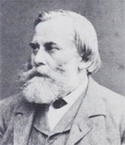
Fig. 4. Dr. Edmund Hartnack (1826-1891).
What is clear from the preceding paragraph is that Freud had a strong preference for the optics produced by Edmund Hartnack. This was a well-placed trust for Hartnack as an innovator (Bradbury, 1967) whose instruments are highly regarded by microscope historians (Moe, 2004). Edmund Hartnack (Fig. 4) was born on April 9, 1826, and died on February 9, 1891. In 1830, Georges Oberhäuser (1798-1868), an uncle of Hartnack, had combined forces in Paris with Trecourt and Bouquet to form a fine instrument-making firm. From about 1835, Oberhäuser worked alone, achieving a reputation for quality. In 1857 he went into partnership with his nephew Edmund Hartnack (Oberhäuser & Hartnack, Place Dauphine 21, Paris).
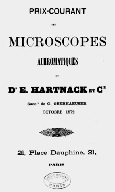
Fig. 5. An October 1872 price list for Hartnack microscopes from the Paris branch.
Hartnack assumed sole control of the firm in 1860 (Fig. 5) and continued the manufacture of microscopes with the same skill and innovation that made his uncle famous. In 1870, as a result of the Franco-Prussian war, Hartnack moved to Potsdam (Fig. 6), leaving the Paris division (Hartnack & Prazmowski, Rue Bonaparte 1, Paris) to his partner Adam Prazmowski (1821-1888), a Polish professor of Mathematics and Astronomy, who had previously worked at the Warsaw Observatory.
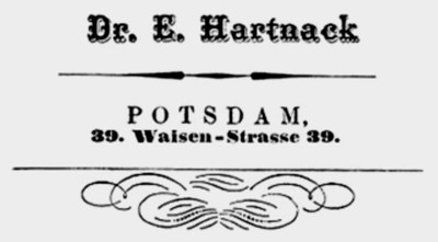
Fig. 6. Hartnack logo from the Potsdam years.
The Parisian branch of the business at some point was renamed Prazmowski Bezu & Hausser, and was eventually taken over by Nachet et Fils in 1898. Hartnack is credited with the first use of water-immersion lenses in the commercial production of microscopes and the adoption of the substage condenser in his later instruments.
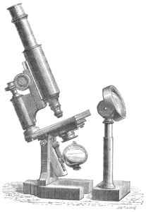
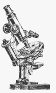
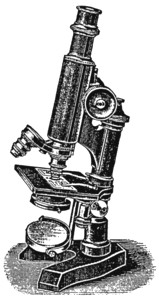
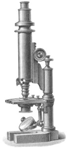
Fig. 7. Various engravings of Hartnack microscopes.
Microscopes by Hartnack (Fig. 7) can be found in several microscope collections (Purtle, 1974; Turner, 1989), including our own (Figs. 8 and 9). We present brief descriptions of two of these Hartnack microscopes which are contemporary with the one that allowed Freud to observe nerve cells in the laboratory and to reach his fundamental histological conclusions.
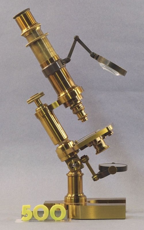
Fig. 8. A Hartnack & Prazmowski microscope from the Paris period
(collection of MdC).
Microscope #500 in the MdC Collection (Fig. 8) Signed E(dmund) Hartnack & A. Prazmowski, Rue Bonaparte 1, Paris. Brass monocular with attached bull’s eye condenser, serial #12460. The horseshoe base is 11.4 cm long by 7.5 cm wide. A 3.8 cm tall pillar carries the adjustable inclination joint. The arm is straight; at its lower end it offers attachment to the 4 cm-long swing arm holding the fork that supports the mirror. The mirror is plano-convex, 3.7 cm in diameter. The stage is 7.7 cm wide and 6.0 cm deep; it retains the original stage clips. A sliding brass plate held in the understage holds the fix aperture diaphragm. The arm is 7.0 cm tall and it carries the fine focus knob on its top. A horizontal arm ending in a ring holds the brass sleeve that supports the microscope body tube. The sleeve supports an articulated arm that holds the bull’s eye condenser used for incident illumination. The body is 10.0 cm tall; it contains a drawtube into which an ocular labeled “1” is inserted. The body is signed “E. Hartnack & A. Prazmowski, Rue Bonaparte 1 Paris.” The original wooden case is 27.7 cm wide, 15.2 cm deep, and 10.2 tall; it retains its original key. A rack inside the box holds two extra oculars, one is marked “3”, and the other “4”. A small drawer attached to the inside of the case contains an all-brass ocular that carries no label. There are also two extra brass objectives, one is labeled “1”, the other is unmarked. A small box covered with green leather and lined with silk is loose inside the case. It measures 7.3 cm wide, 3.1 cm tall, and 4.8 deep. It contains a wooden block with holes for holding three objectives and three diaphragms. Two objectives and two fixed diaphragms are kept in it; there are empty spaces corresponding to the objective and diaphragm mounted on the microscope. The diaphragms are unlabeled; the objectives are inscribed “7” and “9”. The wooden block is inscribed with the number “12460.” Provenance: Librairie Alan Brieux, 48 Rue Jacob, Paris, France. Date: May 1997.
OPTICAL PERFORMANCE: Ocular #3 could not be tested because its field lens is smeared with remnants of a dried up fluid. Accordingly, the following combinations were tested:
Ocular #1 with objective #2 gives a good image with some curvature of field; the field diameter is 770 µm.
Ocular #4 and objective #7 gives a good and flat image; the field diameter is 450 µm.
Ocular #4 and objective #9 gives a faint image; which makes measuring of the field diameter ambiguous.
COMMENTS: This circa 1864 instrument is an early example of the “continental” design. It was made by one of the originators of such style, which later became almost universal in its acceptance.
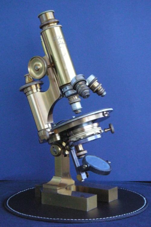
Fig. 9. A Hartnack microscope from the Potsdam period
(collection of LCT).
A later Hartnack model, from the Potsdam branch, is shown in Fig. 9. Details: Engraving on the tube: E. Hartnack Potsdam. Serial no. 25132 (marked on lens box). Ocular set: 2, 3, 4. Objectives: 4, 7, Hom. Imm. No I.
Similar in design to Oberhäuser instruments with a leaded brass, horseshoe-shaped base 12.3 cm long × 9.0 cm wide, the microscope is mounted on a round brass pillar that holds the mirror, round stage, condenser, and body tube. The body-tube moves by rack and pinion and is connected to the limb by a solid brass arm. A vulcanite surface protects the stage (diameter 10.0 cm), and a plano-concave mirror (diameter 4.7 cm) is mounted beneath the condenser. Beneath the stage, which retains the original clips, is a collar for an Abbe illuminator equipped with an iris diaphragm. The eyepieces are Huygenian-style, containing two lenses, and the interchangeable objectives are screwed into the lower end of the turret nose-piece.
The original wooden case measures 21.0×20.0×33.8 cm (W×D×H). A rack with four round openings (diameter 2.35 cm) holds the two extra ocular lenses and an empty brass canister engraved “1/12 E. Hartnack Postdam” on the cap. An accessories drawer (8.0×15.6×3.1 cm) is attached to the upper inside of the case. A box (7.5×5.0×3.3 cm) covered with brown leather and lined with blue silk is attached to the upper inside of the case, opposite from the drawer; it contains a removable wooden receptacle, inscribed “E. Hartnack Potsdam” and the number “25182”, with three sockets (diameter 1.95 cm) for objective lenses and three sockets (diameter 1.05 cm) for diaphragms. The first two sockets are marked “4” and “7”, matching the objectives mounted on the microscope. The third socket is unmarked.
Comments and criticism to the authors are most welcome.
REFERENCES
Bradbury, Savile (1967) The Evolution of the Microscope. Pergamon Press, Oxford, pp. 224-235.
Freud, Sigmund (1877) Beobachtungen über Gestaltung und feineren Bau der als Hoden beschriebenen Lappenorgane des Aals. Sitzungsberichte der kaiserliche Akademie der Wissenschaften (Wien) 75: 419-431.
Freud, Sigmund (1878) Über Spinalganglien und Rückenmark des Petromyzon. Sitzungsberichte der kaiserliche Akademie der Wissenschaften (Wien) 78: 81-167.
Freud, Sigmund (1882) Über den Bau der Nervenfasern und Nervenzellen beim Flusskrebs. Sitzungsberichte der kaiserliche Akademie der Wissenschaften (Wien) 85: 9-46.
Gay, Peter (1988) Freud, A Life for Our Times. Anchor Books Doubleday, New York, 808 pp.
Guttman, Giselher and Inge Scholz-Strasser, eds. (1998) Freud and the Neurosciences – From Brain Research to the Unconscious. Verlag der Österreichischen Academie der Wissenschaften, Vienna, 116 pp.
Moe, Harald (2004) The Story of the Microscope. Rhodos, Denmark, pp. 191-194.
Panek, Richard (2004) The Invisible Universe: Einstein, Freud, and the Search for Hidden Universes. Viking, New York, 258 pp.
Purtle, Helen R., ed. (1974) The Billings Microscope Collection. Armed Forces Institute of Pathology, Washington, D.C.
Ramón y Cajal, Santiago (1897-1904) Textura del Sistema Nervioso del Hombre y de los Vertebrados, volúmenes I-III (original Spanish edition). Moya, Madrid. Histologie du Système Nerveux de l’Homme et des Vertébrés, 2 vols. (French translation by L. Azoulay). Maloine, Paris, 1909-1911. Histology of the Nervous System of Man and Vertebrates (English translation by N. Swanson and L.W. Swanson). Oxford University Press, New York–Oxford, 1995.
Triarhou, Lazaros C. and Manuel del Cerro (1985) Freud’s contributions to Neuroanatomy. Archives of Neurology, 42: 282-287.
Triarhou, Lazaros C. and Manuel del Cerro (1987) The histologist Sigmund Freud and the biology of intracellular motility. Biology of the Cell 61: 111-114.
Turner, Gerard L’E. (1989) The Great Age of the Microscope: The Collection of the Royal Microscopical Society through 150 Years. Adam Hilger, Bristol, p. 210.
Published in the October 2006 edition of Micscape.
Please report any Web problems or offer general comments to the Micscape Editor .
Micscape is the on-line monthly magazine of the Microscopy UK web site at Microscopy-UK
© Onview.net Ltd, Microscopy-UK, and all contributors 1995
onwards. All rights reserved.
Main site is
at www.microscopy-uk.org.uk
with full mirror
at www.microscopy-uk.net
.