 |
 |
 |
 |
| Reducing Lighting
Anomalies in Low-Medium Power Darkfield Illumination Getting the best out of the Abbe condenser By Paul James |
If anyone offered you a $1.00 bottle of Dr Fix's 'cure all' sporting a list of ailments from A to Z then you'd be rather suspicious to say the least, and understandably so. I mention this because the 'cure' I offer here appears at least in microscopist's terms to originate from the same ilk as having a pivotal role in Köhler illumination and in certain Brightfield applications too. Despite this, I hope the reader's mind is an open one, if not only at least to try the method first and then make a judgement for themselves, whatever that turns out to be !
The Problems
Most suitable specimens for darkfield tend to be self contained, isolated structures or the like which enhances the visual effect of this illumination technique. Even so there are times when using low objective powers when the maximum field illuminated is either insufficient to cover the specimen and or that it's peripheral boundary is compromised, as well as prominent features in the specimen by prismatic effects from the simple non achromatically corrected Abbe substage condenser. The condenser can be used without its top lens, but there is a void between these extremes which is the problem. Acquiring an achromatic/aplanatic condenser will no doubt improve the situation BUT the field coverage might be no better or rather less than that of an Abbe.
An Effective Cure.........Ground Glass screens
Of course the beauty of this technique is its cost effectiveness improving both the field width problem as well as the coloured field margin too. If the idea of employing a piece of ground glass in Bright Field seems a little irrational, then its use in Darkfield must appear even more destructive to the image in theory?? However the fact is that it works extremely well. Thus by placing a suitable ground glass screen immediately below the condenser we can broaden and cleanup the DF effect in low power use. Surprisingly, the imagined reduction in contrast does not appear to be of any significance as the images below seem to testify. Visually the imagery so produced only changes in intensity, which is to be expected of course, but how the chromatic artefacts magically disappear is a more complex issue I suspect which fortunately does not have to be understood.
I've illustrated this method by taking images with and without the ground glass screen in DF. The light source for DF comes through annuli and not traditional dark stops. The subject matter is of no importance here save only to illustrate the ground glass's effects. The position of the diffusing screen is best immediately below the substage condenser.
All images below have been sized and cropped. Microscope settings remained identical for each pair of exposures. The image's are effectively 'out of camera'.
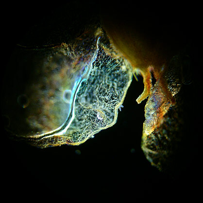
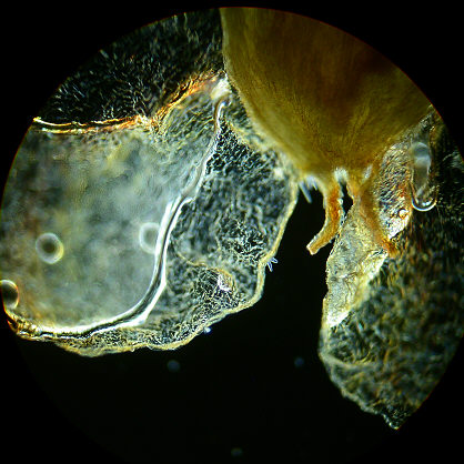
NO SCREEN ...................................................................................GROUND GLASS SCREEN ( 180 grit carborundum )
|
|
|
|
The two images above clearly demonstrate the effects as the diffusing screen removes the colour artefacts extremely well and broadens the field somewhat. The differences in visual intensity between the two where compensated for by using the spot metering facility of the camera on the same part of the specimen. The intensity and form of colour artefacts generated by the Abbe condenser varies of course between condensers, and with the specimen type, its mounting and also objective power.
 |
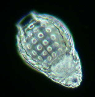 |
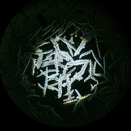
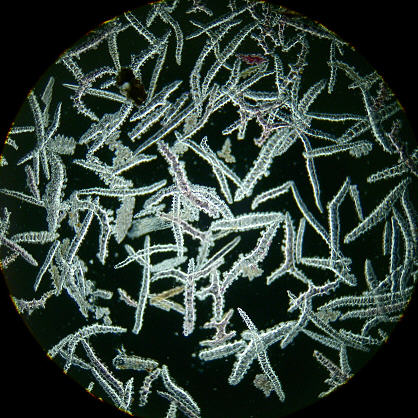
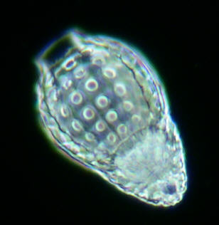
These two images of a Radiolarian have been cropped from a small area from the centre of two medium power DF images with the image right having the diffusing effects of a ground glass sheet exactly as the first examples above. The purpose of showing these is to demonstrate rather more critically that there are no material disadvantages in using a diffusing screen in DF. The exposure levels for the camera were obtained using its spot metering facility which enabled fairly consistent imagery. Notice that all the internal prismatic effects of the specimen's structure are virtually identical in these images. ( Radiolarian. x20 Wild Plan Fluotar objective )
The Darkfield Stop
I also find that when using a traditional DF stop which is a simple circular disc, it can produce overly bright and flooded imagery. An annulus of the type used in Phase Contrast or COL illumination is ideal, more especially if its NA is just beyond that of the objective in use. Aside the distinct advantage of controlling the amount and angle of light that bathes the specimen from an annulus, there is also the benefit of only using that light which is within the peripheral regions of the condenser's optics which suppresses the chromatic artefacts. Thus the annulus permits the output of a thin coherent cone which has greater delicacy of imaging than the standard DF stop, and so having for each objective in use a 'designated' annulus promises the very best DF potential.
Conclusions
The DF coverage with the intervention of the ground glass diffusing screen is modestly increased and therefore of some significance, but it could make all the difference especially in visual use, as well as its effective diminishment of prismatic effects from the Abbe condenser when observing larger specimens in low power. The only observable difference apart from those stated is a slight lightening of the darkground, but this is really of no consequence to the eye or indeed the camera.
The idea of using ground glass screens seems deplorable to some individuals, but the fact is that its use in microscopy is significant and widespread. That diffusion screens work well in both BF and DF in certain circumstances gives credence to the eye and mind 'combo' which have evolved over the millennia under nothing more than diffused lighting from the sky !
Finally I must advise anyone who might be interested in experimenting with diffusion screens to restrict their interests to ground glass only, as semi opaque plastics etc.. have an entirely different effect on light which might at first sight seem suitable.
| All comments welcome by the author Paul James |
Microscopy
UK Front Page
Micscape
Magazine
Article
Library
Please report any Web problems or offer general comments to the Micscape Editor.
Micscape is the on-line monthly magazine of the Microscopy
UK web
site at Microscopy-UK