Using Affordable Technology With Microscopy
'Stacking, Stitching, Slowing'
by Mol Smith Oct. 2013
Rapid Change
Wow! We now live in an Apps, iPad, social media connected world. {Groan}. Sometimes, I just wish to get off the planet to escape the hype, the data-noise, the marketing, the over-non-information. But within the noise, there are some powerful resources by like-minded people which can be used to help exploration and one's individual fascination with microscopy. So, I thought I would start to explore some of that and share it with
you.
Stacking (over-coming narrow depth of field)
No. Not filling shelves in Tesco or Walmart (ASDA in the UK)... photo-stacking: a boon for microscopists with cameras fitted to their microscopes. Try taking a photograph of a micro-organism which is almost flat to the untrained eye, with a fine focus which, even when just twitched just a few micrometres, means flying through the whole organism and out the other side - and you get to understand how difficult it is to produce a 'simple'
photograph of something under an affordable microscope. What you really need is a set of photos at varying depths into the subject and a way of combining only the best, in-focus elements from each image into a composite one.
A diagram helps to explain - sometimes...
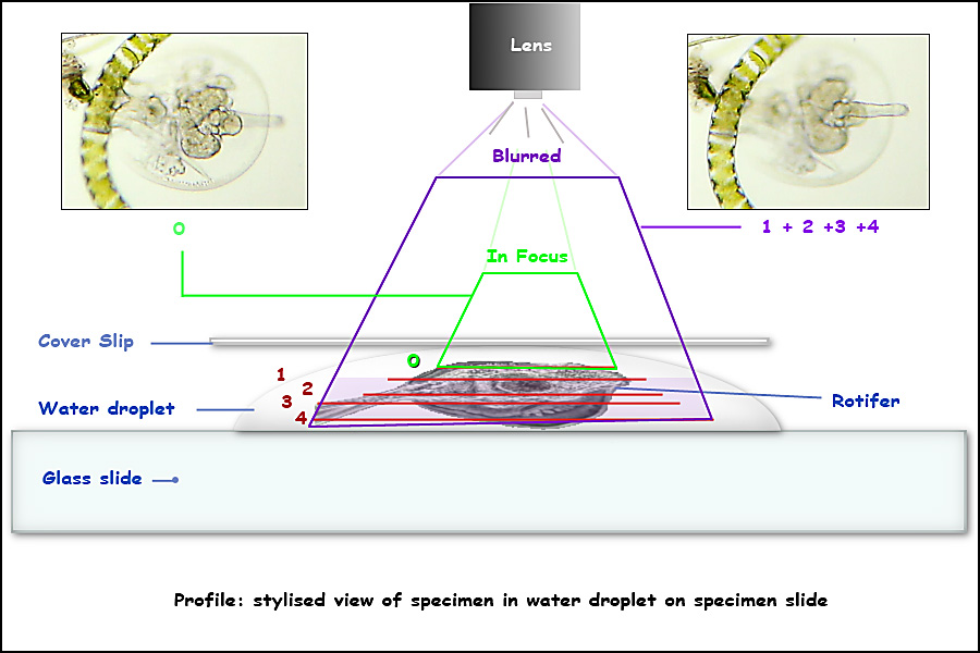
In the diagram above, if you are focused on depth [0] details in depths [1 to 4] will be out of focus. if you focus on depth [3] details in depth [1 to 2] and [4] will be out of focus. When we use our eye to look, we can twitch the fine focus and build up an understanding of the subject's processes mentally, but obtaining a sharp photograph where all depths are in focus is not possible... that is - unless you use some clever software.
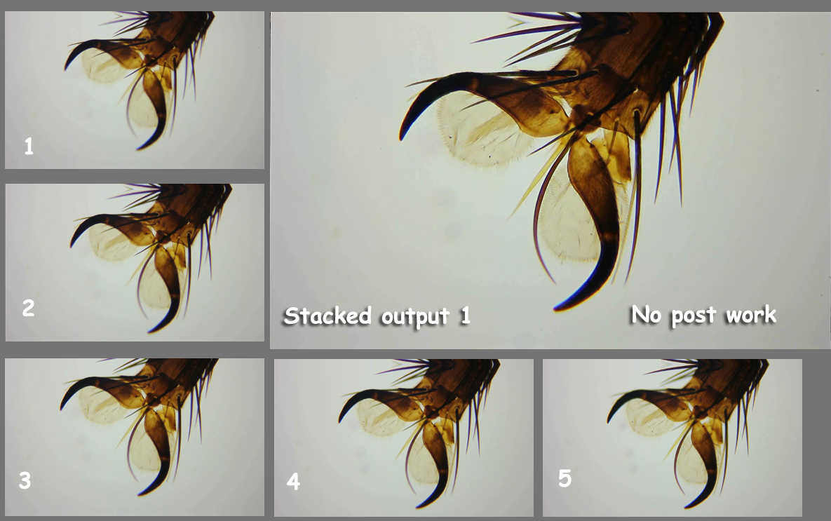
Stacking software enables you to take a series of photographs at different focus levels into the subject and then takes only the in-focus elements in each photo to build a final more-resolved image. Most will allow extracting frames from a video taken through the microscope. I put my Canon 550D on my scope and using 50 fps shutter speed and a time rate of 25 fps video clip rate recorded a second or two as I twitched the fine focus from top detail to bottom detail on a bee claw slide. Using Combine ZP created
by Alan Hadley, I extracted all the frames and stacked them to form a final image. His software allows automatic removal of duplicate frames. Most are. I was left with 6 but deleted one as it looked too blurred to be of real value. This left 5 frames. The frames plus the 'stacked' result is below. You will need to click on each image to see the full size on and the parts in focus in each image in the stack. I could have gone over the final image in Photoshop to brighten it up but I haven't.
The software has a variety of methods for trying to achieve a good 'stacked' photo. I don't wish to explain how to use the software in this short article, only to expose the availability of the technology. There are alternatives to Alan's software but I believe in terms of value, usability and function - it's the best. You might find the interface a bit quirky and need to play with the software for an hour, reading the help files before
realising that Alan has provided you with a lot of powerful tools and functionality to get a great final image. And yes, the software does a good job on transparent specimens too!
Stitching (over-coming limited image area)
Some specimens are too large on higher levels of magnification to be contained in one photograph. But stitching software allows you to scan the subject manually, take photographs of each part and automatically join them together into a composite larger image. Most people are aware of this type of software (often used to create panoramas) but few realise it can be used to 'stitch' together a series of photographs taken down a microscope.
The software should contain a routine to correct lens curvature so the resultant image is correct.
Artificial Slow Motion
Some pond life organisms are extremely fast. I would love to own a high speed time-lapse camera so I could shoot video of cilia movements and other very difficult to resolve high speed processes. By resolve, I mean to see movement in detail. One solution is to use a plug-in for Adobe Premiere Movie Editing software. Premiere is expensive to own but it does allow you to create professional movies of your videos taken at the microscope. A plug-in exists which is fairly affordable which slows down video by calculating
and creating in-between frames - 'tweening'. The plug-in is called Twixtor, licensed by Revision (see references below). Allow me to demonstrate.
In the video below is a rotifer with apparent spinning cilia. We all know, of course - they don't really spin. A great optical illusion. But I would like to slow down the cilia and indeed the mastax (jaws) so I can see the two halves of the mastax come together. Watch the following video to see some of the benefits of using Premiere and Twixtor.
|
|
References
| Stacking Software | Price 1 | Price 2 | Created for |
| Combine ZP | Free | microscopy * recommended | |
| Ł30 annual licence | Ł115 unlimited time licence | microscopy | |
| Deep Sky Tracker | Freeware | Freeware | Astronomy |
| Zerene Stacker | n/a | $89.00 | microscopy |
| Stitching Software | Price | Notes | |
| Autostitch | 399 Euros | Also called Panotour Pro | |
| Panorama Factory | $79.95 |
External Information Sources
Another article comparing Stacking software by Tom Mortimer
TWIXTOR Premiere plug-in ($66.00)