|
Sea Capers, Sea Pickles, Sea Cucumbers and Sea Marrows: A Brief Introduction to Holothuroids by Richard L. Howey, Wyoming, USA |
Holothuroids or sea cucumbers as they are commonly known, as intriguing and remarkably strange as they are, will never win any beauty contest. Some species have extraordinary patterns of coloration, but nonetheless are not creatures that you could cozy up to nor would they make good pets.
Sea cucumbers are echinoderms (“spiny skins”) and some are occasionally described as “hedgehogs of the sea”, but looking at sea cucumbers will make you wonder why they have been placed in the phylum Echinodermata.
They don’t appear to be spiny and they certainly don’t look like hedgehogs. So, we have to remind ourselves that taxonomists are also very weird creatures. Imagine grown people–ostensibly adults–having fist fights over the issue of synonymy of species–not a common event, I admit, but such altercations have happened.
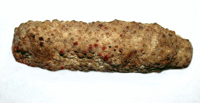
The general body plan is in general–always a necessary qualification when talking about groups of complex organisms–that of an elongated, tubular sack. It can be relatively small, a “sea caper” as in the case of Lissothuria, or a “sea pickle” as in the case of Eupentacta pygmaea which may be only a couple of inches in length. First an image of Lissothuria; this specimen is about 10 mm long and then a Eupentacta which is about 50 mm long.
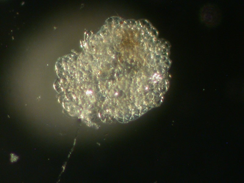
Here, of course, in this preserved specimen, you can’t see any tentacles because they are retracted, so in life it would be a bit bigger. This species is of interest because the dermis, just under the surface, is an almost solid mat of tiny beaded, calcareous skeletal plates–crunchy armor to discourage prey. These structures are called spicules or ossicles and in different species vary greatly in shape, quantity, and size. There are a few types that are sufficiently distinctive so that they can be of help in identification. The ones in Eupentacta are of a fairly common type and consist of small fenestrated clusters of a few spheres or “beads” as can be seen below.
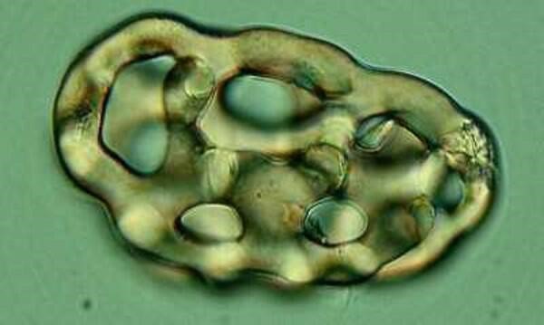
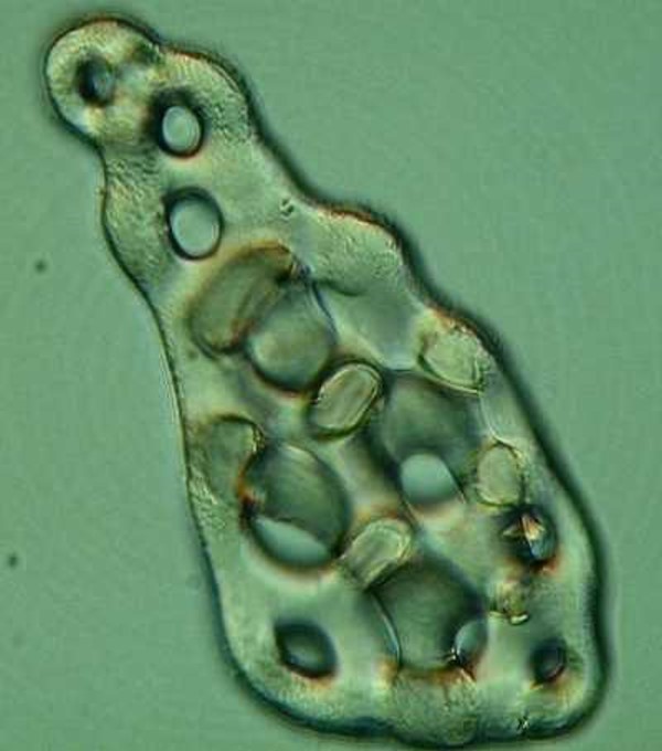
Fenestration simply means that these structures are permeated with small holes or “windows”. (When I was still teaching I had an office on the 4th floor of the Humanities building and when a student would get insolent, I would threaten to defenestrate him or her. (I suspect most of them thought I meant something obscene and those who did understand defenestration couldn’t take me seriously because the window was too small.) Fenestration is crucial for the skeletal structures of these organisms for without these “windows” the weight would be unmanageable; think what a monumental chore it would be for a Heterocentrotus mammilatus to move at all if its spines were solid.
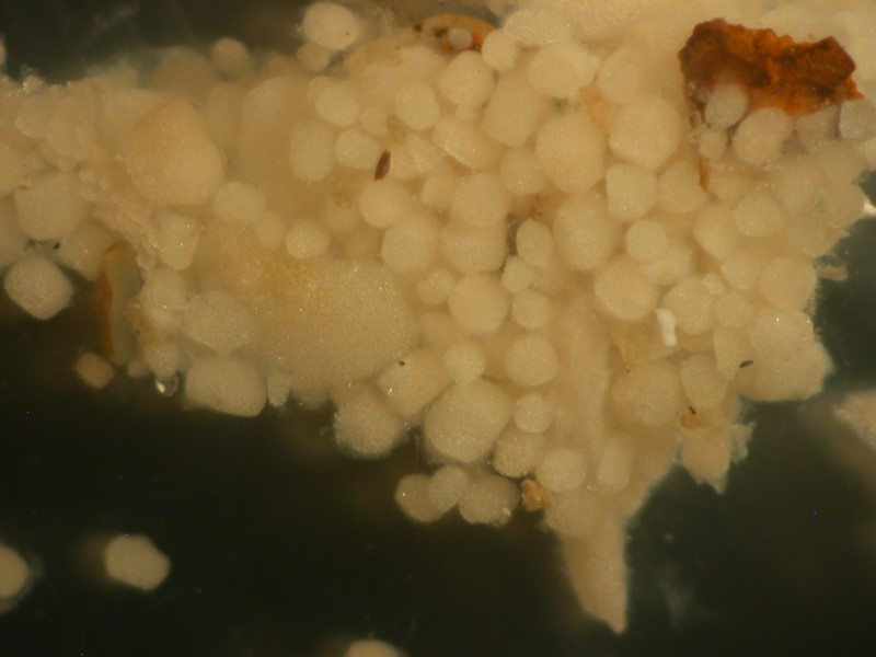
Chirodota and closely related genera have wheel-shaped spicules.
These occur in small pockets distributed irregularly in the surface tissue of the organisms. The first image shows a group of packets in the skin and the second image shows a single packet.
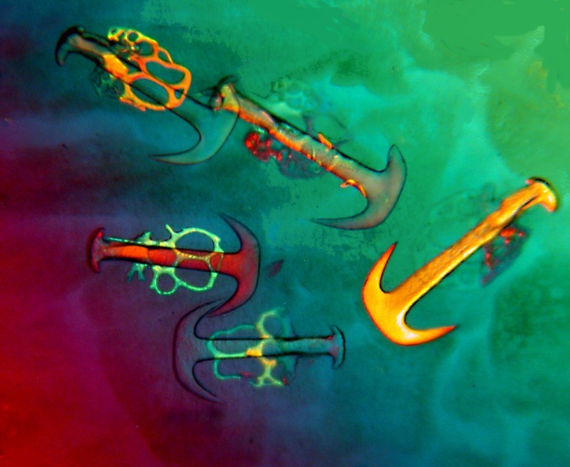
Some species of Synapta also have distinctive ossicles which consists of fenestrated plates and anchors.
A bit later we’ll talk about the possible function of such structures.
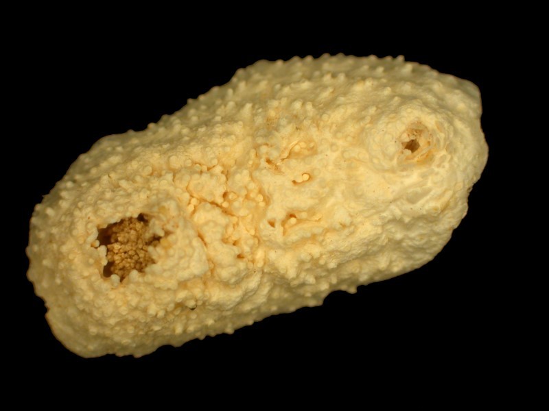
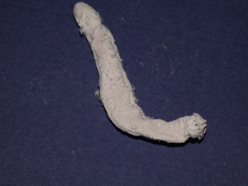
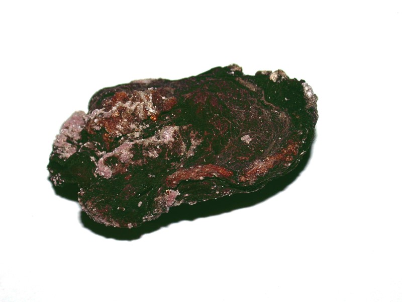
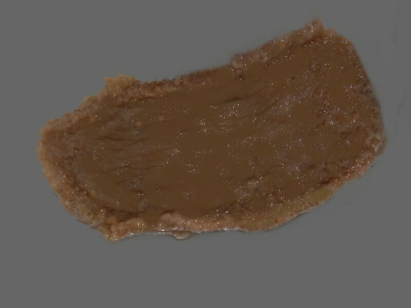
The “standard” sea cucumber has a considerable variation in size range. Cucumaria salma, for example, is, in its contracted state about 3 inches and has the shape of a slightly ovoid sphere. Unfortunately, the dried specimen which I have is greatly distorted.
In order to give you a glimpse of what the appearance is when living, check this link.
The pattern is typical of this species.
C. frondosa is the species often available from biological supply houses for classroom study and is a fairly typical paradigm if there is such a thing for a sea cucumber. This species is often 6 or 8 inches long.
Then, there are species which are over 6 feet long!
As I mentioned earlier, no matter how colorful, these are really not very attractive creatures, but are fascinating nonetheless.
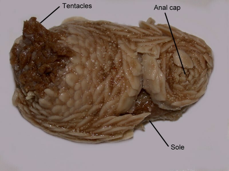
The majority of holothuroids crawl along the bottom and these forms are usually described as having a bilateral external symmetry consisting of a ventral area with three sets of rows of tube feet (this area being designated the “trivium”) and the dorsal area consisting of two sets or rows of tube feet (designated as the “bivium”). Naturally there are modifications and exceptions. The genus Thyone has tube feet scattered across the whole body surface.
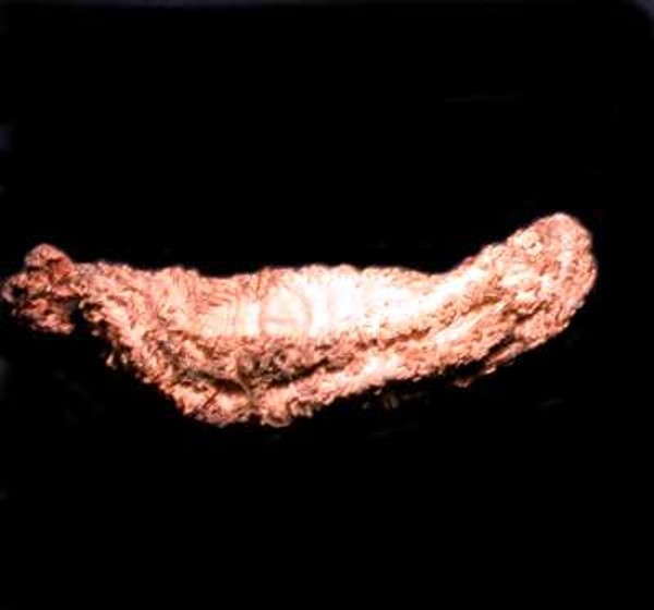
Psolus has scales on the dorsal aspect and a flattened “foot” as its ventral surface. First, the dorsal aspect with the scales and then the “foot”.
Other genera, such as Synapta, are burrowers and some form a “U”-shaped tube with the tentacles positioned at one end and the anus at the other. Yet others have an essentially vertical burrow with the tentacles, anus, and gonopore all located at the anterior end. And then, there are a few species of holothuroids that are pelagic and look like some ultra-bizarre jellyfish.
While many holothuroids have an external bilateral symmetry, the internal symmetry is almost always pentamerous (that is, based on the number 5 like any good, self-respecting starfish, except for those that have 4 arms or 6 or 7 or 12 or 31 arms). So, in a sense, we might say that there is a kind of morphological schizophrenia in virtually all holothuroids, namely that in spite of their external configuration, they tend to be pentamerous internally. Exactly how this is the case is a complex topic which will have to wait for a future essay.
The external features which are most obviously significant are the tentacles, the dermal ossicles, and the podia or tube feet.
Since, we’ve already seen a couple of examples of dermal ossicles (=spicules), let’s start there. I already mentioned that Eupentacta pygmaea has thousands upon thousands of these like a suit of chain mail armor which is a rather good analog since such armor was also fenestrated to reduce weight and heat. Some species of Chirodota, as we have seen, have little isolated packets of spicules which may contain a hundred or so “wheels”. In yet other forms, the spicules are sparse and randomly dispersed and then there are some genera which apparently have no dermal ossicles or, if they do, they have been reduced to extremely tiny, amorphous, birefringent bits, rather like micro-micro sand grains. At this point the question inevitably arises: What function do these ossicles serve?
Here, we need to digress a bit and consider briefly the issue of calcareous dermal structures in echinoderms in general. The most familiar examples for the majority of people are the asteroids or starfish. These amazing creatures capture the attention of both the young and the old and the attraction they exert over us leads to our displaying them everywhere from our own cabinets of curiosities to nets hung in tropical bistros. The plates and spines of asteroids show, as you would expect, a wide variety of size and shape. An instructive exercise is to take a small to medium-sized specimen of a common starfish, such as, Pisaster and subject it to the Torquemadian tortures of household bleach.
Hopefully the specimen is already preserved, but it does raise the question: Is there a “humane” way to kill starfish? I admit that such a question is not at the top of my list of ethical considerations, but being an eccentric through and through, I can’t help but consider such issues on occasion.
The first time one carries out this procedure, it is almost impossible not to be astonished at the huge number of plates and, in some species, thorny, spine-like strictures as well.
It would seem that all of this baggage would make these creatures ponderous and ungainly until one considers the fenestrated character of these structures which results in a considerable weight reduction. I suppose if you’re radically calcium-deficient and really like crunchy stuff, you might consider munching on starfish but, even then, you have to be careful about which species you select for an appetizer. Acanthaster plancii, the “Crown-of-Thorns” starfish exudes a toxin from its spines thus further narrowing its range of predators.
However, imagine a very large snail, the Giant Triton (Charonia tritonis) snacking on this starfish. Unfortunately, because it has a very impressive shell, it has been severely over-collected thus allowing Acanthaster to overpopulate and, as it eats coral, it has done great damage to tropical reefs and their inhabitants which depend on the reefs.
For some reason, the Giant Triton is undeterred by the toxin or the spines, making a main course out of this bristly creature. Mother Nature has a very strange sense of humor.
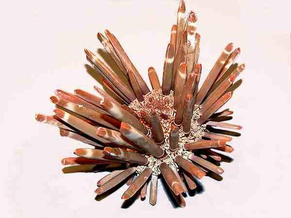
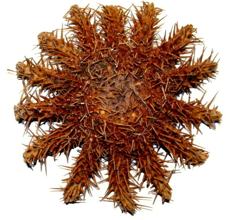
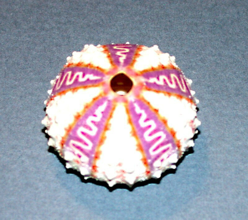
If we take a glimpse at the echinoids (sea urchins), we again find a variety of types of armor, some it very formidable indeed. The internal organs are encased in a shell or “test” which consists of a series of calcareous plates fused together and many of these tests are quite lovely objects and some nature enthusiasts collect them. There are two basic types of echinoids–the regular and the irregular. the regular ones have a test that is nearly spherical or dome-shaped with the bottom part truncated where there is a roughly circular opening for the teeth. Some are quite small (less than an inch in diameter) and are relatively fragile whereas others have a diameter of 5 or 6 inches. Below are 2 lovely examples; the first is Coelopleurus exquisitus from New Caledonia and the second is Stereocidaris grandis from the Philippines.
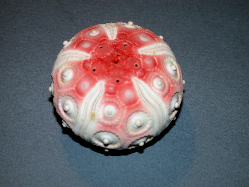
As you can see, the test itself provides a considerable degree of protection to the internal, soft parts of the organism; however, it doesn’t end there. All those little “bumps’ on the surface of the test support spines ranging from the tiny acicular (needle-like) ones of Mespilia
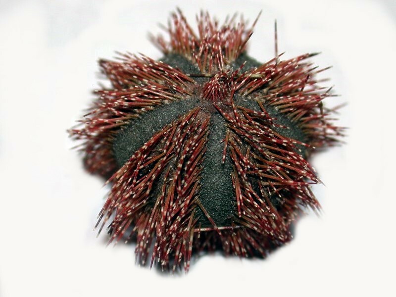
to the “pencils” of Heterocentrotus mammilatus.
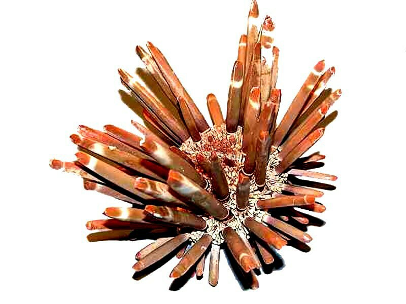
In addition, there are species of Diadema which possess spines over a foot long and which are hollow and have venomous glands.
So, safe from predators, you’re thinking. Well, not quite, If you’ve ever observed very young human children, you will very likely have observed how strong a gustatory disposition they have–they want to taste everything that crosses their path. so, in some cultures, the internal parts of sea urchins are regarded as a delicacy and there are also certain sea birds which have devised a clever strategy for extracting the “tasty”, meaty parts. They dive down, grasp the urchin with their talons, fly up over a rocky spot along the beach and then drop it, letting it smash open on the rocks and then they can feed on the morsels. Nonetheless, in general, echinoids are, well-protected. Crinoids (“sea lilies”) and brittle stars also have lots of crunchy bits in the way of plates, spines, and spicules and so many can also be described as fairly well-armored.
Sea cucumbers, however, pose some puzzles in this regard. With the exception of a small number of species, such as Psolus which has dorsal scales or Eupentacta which has a dense dermal layer of spicules, Holothuroids are not very well armored and some species seem to have no dermal ossicles at all or, at best, none of any significant size. It is intriguing that in a single phylum we find such variation and the echinoderms are not unique in this regard. Some mollusks have heavy, massive shells that afford a high degree of protection (except from shell collectors) whereas others, such as the pteropods (“sea butterflies”) and octopuses and squid have at most the vestige of a shell. Nature constantly evolutionarily experiments and I suspect that we humans are one of her less successful experiments–big brains, but an inability to use them well.
All comments to the author Richard Howey are welcomed.
Editor's note: Visit Richard Howey's new website at http://rhowey.googlepages.com/home where he plans to share aspects of his wide interests.
Microscopy UK Front
Page
Micscape
Magazine
Article
Library
Published in the October 2016 edition of Micscape Magazine.
Please report any Web problems or offer general comments to the Micscape Editor .
Micscape is the on-line monthly magazine of the Microscopy UK website at Microscopy-UK .
©
Onview.net Ltd, Microscopy-UK, and all contributors 1995
onwards. All rights reserved.
Main site is at
www.microscopy-uk.org.uk .