Notes on the birefringence of ice or its appearance between crossed polars.
by David Walker, UK
In earlier articles I've shared my trials of using Peltier plates as cold stages for both a compound and stereo microscope. A particular aim was to explore the formation of ice, either constrained under a coverslip or uncovered to form frost in the comfort of a warm room. In the May 2018 article where I described these ice studies I remarked that "Ice is not birefringent for polarised light studies so variations on darkfield were best". Tut-tut, this was incorrect but fortunately readers haven't to date been beating a path to my door pointing out my misunderstanding of the observations. I only appreciated this error when splendid imagery of ice showing birefringent colours under crossed polars was shared on the You Tube 'Amateur Microscopy' group page (see Acknowledgements).
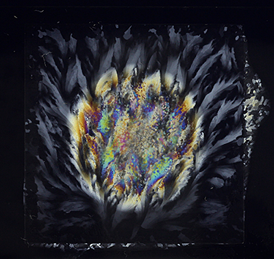
Quantitative polarised light microscopy is an area I still struggle with but have recently tried some simple studies to appreciate better how ice does behave under crossed polars with the key parameter varied being its thickness. The image below shows a cavity microscope slide filled with water with a 22 mm square coverslip. The water film extends to the slip edges so there is both a thin water film and the increasing depth in the cavity. It was placed in the freezer section of the fridge until frozen and quickly photographed on a light box between crossed polars.
Cavity slide filled with water to square coverslip edge. Crossed polars, light box and macro DSLR setup.
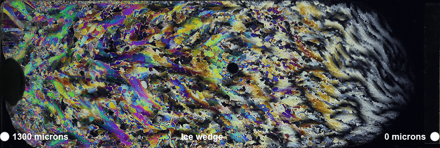
Ice wedge created from two 3 x 1 inch microscope slides, taped touching one end and a spacer of ca. 1.3 mm the other end.
Crossed polars, light box and macro DSLR setup.
Ice is weakly birefringent with the difference between refractive indices of the extraordinary and ordinary rays Δn = 0.004 compared with for example calcite of -0.172 (Wikipedia 'Birefringence' entry values). If a Michel-Lévy birefringence chart is inspected* for the ice value of 0.004, for a sample thickness of 30 microns (thin rock section standard) and below, the interference colours are in the first order deep greys to black; I overlooked them or were barely visible with the thin water samples I had been using in the Peltier plate studies. Extrapolating beyond the chart boundaries, it requires much thicker samples than those typically used on a microscope slide to exhibit colours. The greyscales are visible on the flat outer edges of the cavity slide above where the water thickness was probably thicker than used before. As the water thickness increases into the concavity, interference colours are seen.
The ice wedge slide above shows a similar result as thickness increases from right to left (the water may have melted at its very thinnest portion).
(*Zeiss have a splendid chart to download as a 7 page Acrobat file with accompanying notes on its use and underlying theory.)
Comments to the author David Walker are welcomed.
Resources
A Google search reveals a wealth of resources on ice birefringence and its value to scientists and also a basis for practical science studies at home. Two example resources are below.
Thin Ice: Looking at Birefringence A six minute YouTube video in NASA's Launchpad series illustrating the value of birefringence in ice studies. 'Thin' in this context is not the typical 30 microns of microslide thin rock sections but slabs some cm thick first cut with a chainsaw!
Birefringence of Homemade Ice. Or how to manipulate ice thickness for coloursplosions... An attractively presented web page on Brian Robin's splendid website illustrating practical approaches at home for exploring ice between crossed polars. His other ice projects also include Watching Water Freeze (timelapse) and That Icy Birefringent Palette.
Acknowledgements
Thank you to the Facebook contributor Loes Modderman who regularly shares striking images of her crossed polar studies of chemicals and other materials. One set showing a Petri dish of water frozen in the freezer prompted my own freezer studies after realising ice is indeed birefringent.
Published in the October 2018 edition of Micscape.
Please report any Web problems or offer general comments to the Micscape Editor .
Micscape is the on-line monthly magazine of the Microscopy UK web site at Microscopy-UK
©
Onview.net Ltd, Microscopy-UK, and all contributors 1995
onwards. All rights reserved.
Main site is at
www.microscopy-uk.org.uk.