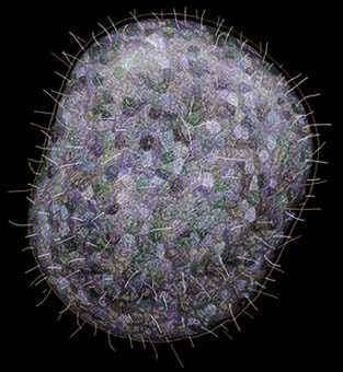
An artist's
impression of Trichoplax adhaerens prepared by Wim van
Egmond using Adobe Photoshop. |
 |
A Weird Wee Beastie: Trichoplax adhaerens
by Richard L. Howey, Wyoming, US
Living nearly 1000 miles from the nearest seashore, I have
little access to marine specimens. Last year, through the
generosity of the proprietors of a local pet shop, I procured
detritus samples from their marine aquaria (see
footnote 1). I was interested primarily in looking for marine
protozoa and was gratified to find a number of fascinating and
elegant types of protozoa. I also noticed what appeared to be a
number of pinkish "deposits" which appeared to be some
kind of odd inorganic, undistinguished something or other. For
several weeks, I was too fascinated with the marine protozoa to
take any further notice. Then, one afternoon, I observed one of
these "deposits" moving. I was looking at sample in a
small culture dish using a binocular zoom dissecting microscope
set on 20x. I immediately zoomed to 40x and observed an
amoeboid-like movement. Initially, I was convinced that I had
found a large and rather strange marine amoeba with a pinkish
tinge.
An artist's
impression of Trichoplax adhaerens prepared by Wim van
Egmond using Adobe Photoshop. |
|
As often happens, I was interrupted and didn't get back to
observing this sample for over a week. Upon resuming my
observations, I found something very odd indeed. The organisms
now looked rather like plasmodial slime molds. They were
"strung out" in unusual configurations which appeared
to be an amoeba with a thin protoplasmic bridge connecting to
another amoeba-like form with yet another thin protoplasmic
bridge connecting to yet another amoeba-like form; yet all of
this comprised a single organism. I had never seen anything
behave in such a bizarre manner. Stranger still, was that there
were folds along some of the edges and sometimes within one or
more of the masses of the amoeba-like parts. These latter folds
seem to project upwards. I took a transfer pipet and tried to
move some of the organisms to a slide to look at them more
closely. Unfortunately, this caused the organisms to come apart
and they were very difficult to get into the pipets, since they
seemed to be glued to the bottom of the culture dish, thus
earning their species name, adhaerens.
I had taken an old aquarium and thrown in
some of the original samples (which contained some filamentous
algal forms) and hoped that this would provide a plentiful supply
of these "weird wee beasties." In a few weeks, it did
and I was able to take samples from the sides of the aquarium and
transfer them to slides and small dishes with minimal damage to
the organisms. However, the first attempts to use slides were not
very successful. The surface tension of the drop would tear the
organisms apart. I finally decided to fit up one of my
microscopes with some old, inexpensive objectives and use them as
water immersion lenses. This allowed me to set the culture dish
on the stage and observe the organisms without transferring or
disturbing them. This led to a great surprise—under 400x, I
observed that the surface of the creatures was covered with
flagella! A flagellated amoeba! I had never heard of such a thing
and was fairly certain that this was not a protozoan at all (see footnote 2).
With much fussing and fidgeting, I was able to get some
video-tape of the organism through the microscope. I then took
the video-tape up to three of my colleagues in the Zoology
department. Two of them are cell-biologists (one of whom is a
specialist in protozoa) and the third is an invertebrate
zoologist who is also a specialist in slime molds. One of them
was initially convinced that it was indeed a large amoeba,
another conjectured that it might be an early stage of a sponge
or a coelenterate or more likely, in his view, a very primitive
non-segmented worm. So, no help from these guys. Off and on for
the next six or eight weeks, I browsed in the library in books on
the lower invertebrates, protozoa, algae, and everything I could
put my hands on that might help me identify this creature. Then
one evening I came across a drawing that looked very much like my
beastie. It was identified as Trichoplax adhaerens
and the drawing also looked like a large amoeba, but the
description reported that Trichoplax is the most
primitive multi-cellular animal known and has the smallest amount
of DNA of any animal ever sequenced. There are reports of another
organism in this group, Treptoplax reptans, but most of
those who know this animal think that the second organism
described is really Trichoplax. In any case, this
organism has a whole phylum to itself, viz., Placozoa.
I was almost certain that this was the right organism, but I
still was puzzled about the odd "strung out" forms
which I had observed and video-taped. So, back to the library.
Now that I had the name, I could check the literature and find
other articles which might confirm my strong intuition.
Eventually I found thirty-four articles, a book chapter, and
three short films on Trichoplax. (Let me say before
proceeding with my ramblings, that I did indeed find an article
on the "strung out" forms and was confirmed in my
opinion that the organism I had was indeed Trichoplax.)
So, now permit me to say something about the discovery and
history of the research on this wonderfully odd animal.
Trichoplax was first described by F.E. Schulze, a German
scientist, in 1883. For a while it was thought to be the planula
stage of a hydromedusan and that theory has been resurrected on
at least two occasions since Schulze. At first there was a good
bit of excitement, but then gradually the view that it was a
planula was accepted and Trichoplax was largely
forgotten. This planula-view was published in an article in 1912.
The next article which I have located appears fifty-four years
later in 1966 again in Germany and then in the 1971, there
appeared the first of many important and definitive studies on Trichoplax
published by Karl Grell, the distinguished protozoologist and
director of the Institute for Zoology at Tübingen. Grell was
also responsible for the three short films on various aspects of Trichoplax.
Trichoplax is an interesting organism to study, because
it is one of those "missing links" that provides some
hints about the evolution of some of the metazoa. It has only
three cell layers and purportedly only four different kinds of
cells. However, it raises interesting questions, some of which
are still not completely answered. How it feeds is still
something of a perplexity. It seems that it absorbs its nutrition
through the ventral surface and probably feeds on algae. I have
observed it draped on pieces of a filamentous alga. In its
immediate vicinity, the contents of the algal cells appear to
have been digested leaving only the cellulose envelope of the
filament.
The extraordinary shapes which it can assume are apparently
accounted for my the middle layer of "fibrous" cells
which seem to provide both a degree of support and also allow for
its radical alterations in form. A student whom I assisted with a
project on Trichoplax was able to show that when the
cells are disassociated; they will regroup, but not necessarily
according to the organism from which they came. He used two vital
stains—red and toluidine blue. When the cells reaggregated
some of the specimens contained cells that were red and others
that were blue.
Trichoplax is an organism that requires a good deal of
patience to study, but it well worth the time and effort.
Apparently not a great deal is known about the ecology of this
organism. Grell got his samples from algal collections that a
colleague of his brought back from the Red Sea. The samples which
I got from the pet shop aquaria are apparently also from tropical
locations. However, Trichoplax seems quite adaptable
judging from the neglect under which it thrived in my cultures,
so it may well occur in areas in which the water is considerable
colder. I suspect that its distribution may be fairly wide-spread
and than it simply has not been looked for in very many
environments. Interestingly most of the reports, other than
Grell's, have come from investigators who happened to notice it
in aquaria. Once it is established in a culture, it is quite
noticeable as small white clumps or stringy blobs on the side of
the aquarium. I have taken to using small fish bowls that hold
about a gallon of water and when the Trichoplax become
abundant in one jar, I start two or three others to ensure a
constant supply. I keep the jars in a window where there is
abundant indirect light. It is important to make sure that algae
will also grow in the culture jars and I usually add 6 or 8
boiled wheat grains to provide food for small organisms that the Trichoplax
may or may not feed on. I use standard marine salts which I
purchase and make up a standard solution with artesian or
distilled water. Trichoplax seems able to tolerate a
range of salinity and so the proportions don't seem critical. I
do monitor the specific gravity of the solution and try to keep
it fairly constant.
When Trichoplax become very abundant in a jar, their
population, for some unknown reasons, starts to decline. For this
reason, it is important to try to keep several cultures going at
once with periodic sub-culturing. When the organisms are healthy,
they have a light rose color. The source of this pigmentation is
unknown. Trichoplax reproduces in three ways: 1) by
binary fission (and this probably takes place when the organisms
are "strung out", 2) by budding, i.e., a spherical body
forms on the dorsal surface and eventually separates off, and 3)
Grell reports the existence of ova, although no sperm cells have
been identified.
Another fascinating aspect of this creature is its ability to
regenerate. As I mentioned above, the organism is often damaged
in the process of transferring it to a culture dish for
examination. However, this damage can be repaired and the
resulting smaller pieces continue to behave as before. Obviously,
there is a minimum amount of material which must be available for
this repair process to take place. In some instances, the
organism seems to just dissipate, but in other instances, after
some days, one may notice a number of very small Trichoplax
in the culture dish.
Another oddity of Trichoplax is that while it is
frequently found either along the bottom or the sides of dishes
or aquaria, I have found some cultures in which significant
numbers of the organism can be found floating on the surface of
the water.
Finally, Trichoplax is an exciting organism to observe,
because it has an odd set of both structural and behavioral
characteristics. When it is "strung out", one can
observe protoplasmic streaming in the "bridges" between
the main masses. There are strange "folds" which
develop along the edges and the dorsal surface of creature whose
function is largely unknown. Along the edges of the organism are
very distinct "shiny spheres". To observe the amoeboid
movement and, at the same time, see the surface covered with
flagella is a remarkable phenomenon. Trichoplax is truly
one of nature's oddities.
Footnotes
1) I want to thank Rick and Jennie Lawrence of Peaceable
Kingdom Pet Shop in Laramie, Wyoming for their generosity and
patience in supplying me samples. Return to
article.
2) Actually there are some protozoa that have both amoeboid and
flagellated forms, but usually only one or two flagella and the
organisms are quite small. Return to article.
Editor's notes:
Comments to author Richard Howey are welcomed.
First published off-line in the Manchester Microscopical and Natural History Society newsletter Micro Miscellanea.
Published in October 1998 Micscape Magazine.
Please report any Web problems
or offer general comments to the Micscape Editor,
via the contact on current Micscape Index.
Micscape is the on-line monthly
magazine of the Microscopy UK web
site at Microscopy-UK