| A look at a transverse section of a stem of a non woody plant such as Ranunculus reveals a variety of tissues. How do these various tissues contribute to keeping the plant upright? | 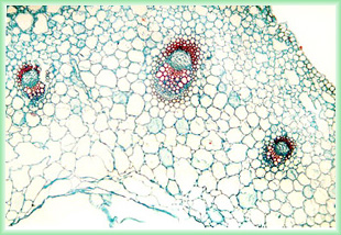 |
| Figure 1 TS Ranuculus stem |
How do non woody plants stay upright?
by Anne Bruce
| A look at a transverse section of a stem of a non woody plant such as Ranunculus reveals a variety of tissues. How do these various tissues contribute to keeping the plant upright? | |
| Figure 1 TS Ranuculus stem |
As can be seen from Figure 1 the tissues comprise an outer epidermis (made up of a single layer of closely packed cells), vascular bundles and parenchyma. Parenchyma cells, which make up the bulk of the stem, are thin walled with large vacuoles. In leaves and stems of seedlings and small plants it is the water content of these cells that holds the plant erect.
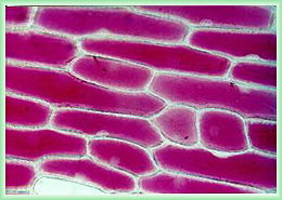
A plant cell is different from an animal cell in that it has a cellulose cell wall and also in that it contains a large sap filled vacuole. If plant cells are placed in a solution which has a weaker solute concentration than the cell sap, then water enters the cell vacuole by osmosis. This causes the protoplast to be pushed against the cell wall exerting pressure against it and preventing the cell from bursting. Cells in this state are said to be turgid. It is this cell turgor that gives the plant support. If however, plant cells are surrounded by a solution with a stronger solute concentration than the cell sap, then the vacuole loses water and as a result there is no hydrostatic pressure acting against the cell wall, such cells are said to be flaccid or plasmolysed. Even those of us who lack green fingers are aware that a drooping, wilting plant has not been watered; once the necessary watering has been carried out the stems and leaves very quickly revert to their erect state as parenchyma cells become turgid.
Turgid and plasmolysed cells can be seen when pieces of the thin membrane from a red onion are placed in different solutions of different concentrations.
 |
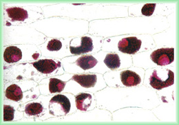 |
| Figure 2 Turgid cells | Figure 3 Plasmolysed cells |
While turgor and osmosis play an important role, other factors are also involved. When transverse sections of dicotyledons stems are observed, vascular tissue arranged in vascular bundles can be seen situated near the periphery of the stem.
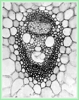 |
Vascular tissue is involved with the transport of material. The vascular bundles are made up of phloem (closest to the outside), xylem, with cambium between. Phloem is a specialised form of parenchyma, made up of sieve elements and companion cells, whose function is the transport of materials produced during photosynthesis. Fibres within the phloem tissue help in supporting the plant. | |
| Figure 4 Vascular bundle |
| Xylem, which does not contain living material, transports water and dissolved mineral salts from the roots and also plays an important role in support. Xylem tissue is made up of tracheary elements, which conduct water and dissolved minerals, together with thick non conducting fibres. The hollow tubes of xylem tissue are usually strengthened with lignin, the woody material we are familiar with in trees and shrubs. The strengthening is in the form of hoops or spirals of lignin as can be seen in the following figure showing lignified xylem vessels in a transverse section of rhubarb (Rheum) | 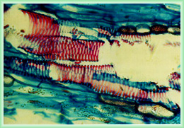 |
| Figure 5 Rheum Xylem vessels showing lignification |
Vascular bundles in dicotyledons can be seen to be arranged near to the outside of the stem; often too, the stem may be hollow. This cylindrical formation provides greater strength than a solid structure of the same weight. Vascular arrangement in the root is different, phloem and xylem are found in a central stele.
| This can be seen in Figure 6 showing a transverse section through a Ranunculus root. The xylem vessels forming the X shaped structure, with phloem inbetween the arms of the X. The vascular tissue is enclosed by a layer of cells called the endodermis. This central arrangement of vascular tissue provides strength against the pulling forces that the root may be subjected to. This same arrangement can also sometimes be seen in the stems of aquatic plants which are subjected to different forces than the stems of their terrestrial relations. | 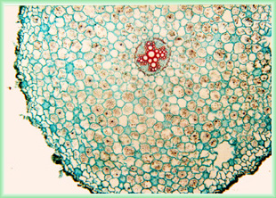 |
| Figure 6 TS Ranuculus root |
Parenchyma, phloem and xylem are not the only tissues visible in a stem cross section; collenchyma often located in the outer tissue has thick cell walls and is relatively pliable, it can play a significant part in the support of shoots but is seldom found in roots. It is often found at the angles of the stem as can be seen in the following picture of a transverse section of a Lamium stem.
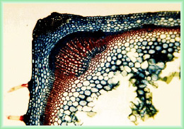 |
| Figure 7 TS Lamium stem |
The cell walls of collenchyma cells are rich in pectic substances; fibrils within the cell wall are arranged such that the cell wall has great plasticity, letting the cells to deform without damage and so allowing the stem to bend without breaking. Often scattered among the cells of the tissues mentioned is a further type of cell, sclerenchyma. This tissue is characterised by having a secondary cell wall (inside the primary cell wall) which gives the cell elasticity, allowing it to return to its original shape after being bent. The angle of the fibrils within the secondary cell wall determines the stiffness of the cell. The arrangement of the fibrils is often varied to confer strength and stability against a range of forces.
Comments to the author Anne Bruce are welcomed.
All Material Copyright © Anne Bruce
Published in the October 1999 edition of Micscape Magazine.
Please report any
Web problems or offer general comments to the Micscape Editor,
via the contact on current Micscape Index.
Micscape is the
on-line monthly magazine of the Microscopy UK web
site at Microscopy-UK