Speaking of cells

  by M. Halit Umar, The Netherlands

.

 
When my daughter Arzu was about 8 years old, she once asked me: 'Papa, what is a cell, actually? Is an egg a cell? Is this apple a cell?

Put yourself in a similar position for a moment and try to find a satisfying explanation to such perceptive queries from a child. Since then, this rare experience continued to occupy my mind. What do we mean when we utter the word 'cell'? What do we really know about the cell? How can we use simple words to make ourselves understood even by a child?

Almost all cells—in biology, of course—are so small, that we cannot see them with an unaided eye. Nowadays, we have instruments through which we can see increasingly finer details of once invisible things. The earlier microscopists successfully founded our basic knowledge during the last centennia, and established the scientific disciplines called microscopy, cytology, cell biology, microscopic anatomy, morphology, histology, histopathology, etc. We can now enjoy the fact that instruments such as the light microscope are readily available and many good models are affordable to microscopy enthusiasts. This allows us to investigate various life forms in extremely variable conditions, all of which have but one thing in common: the cell, a biological unit of living beings.

The aim of this article is to simply present some aspects of living organisms which are observable through various kinds of microscope. So let's explore some basic concepts of cells in order to appreciate their beauty and function.

.

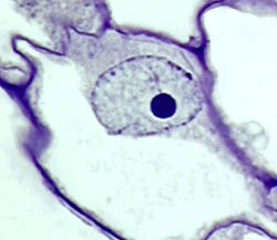
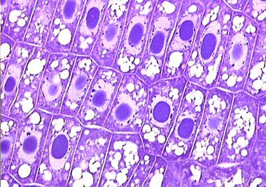
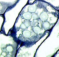
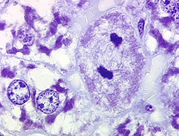
This plant cell is surrounded by at least four other similar type of cells and has a cellulosic cell wall stained dark blue. It contains a large vacuole in which lightly stained granular matter is visible. The nucleus lies at the upper right  and is vesicular (baloon-like). Its nuclear membrane is dotted by darkly stained material called chromatine on which the genetic material is embedded. The nucleus is apparently not empty and contains a moderately well-stained network. But the nucleolus, the dark-blue stained, massive, spherical structure, is the most prominent part of the nucleus. The stained portion around the nucleus is the cytoplasm. The cells illustrated are from a potato, Solanum tuberosum. A very thin section, 2 µm, Toluidine blue (1% aqueous solution) staining.
.

In contrast to its microscopic size, a cell houses an incredible amount of genetic information within its delimiting cell membrane. Inherited genetic information is the driving force of all cell functions such as productions, secretions, excretions and other specilized performances of the cell and biological/chemical activities occur within the protoplasm.

New cells originate from and are identical to a parent cell; the newly born life is comparable with the parent cell in all respects; they can absorb and produce its own substances and may engulf particles. They normally reproduce and replicate, they age and die. The spectrum of the cell's accomplishments are incredible! Their morphological and functional harmony and improvements during evolution are just astonishing. Cells without nuclei (Prokaryotes), cells with nuclei (Eukaryotes), cells with or without mitochondria, cells producing types of a green pigment, the chlorophyll, and capable of photosynthesis; cells making unicellular life forms, cells attached together for a living in groups to form Colonial organisms, from which and then multicellular life forms gradually evolved, as if a step by step development, but in reality only by a gradual evolution taking place in geological time scale; cells forming various tissues after a process known as cell differentiation; and cells making fungi, plants, and animals; cells living in water or on Earth, in an endless, ever increasing variety of niches...

The ultimate achievement of cell development is characterized by the very recent emergence of an animal species characterized by the significant abilities of perceiving, learning, speaking, remembering, judging and decision making; this animal is called man.

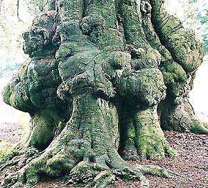
Cells, cells, cells... The building blocks of life. That's right! All living beings are made of cells, without exception; neither for bacteria which first appeared several billions of years ago nor for Homo sapiens sapiens like you and I.

~~~~~

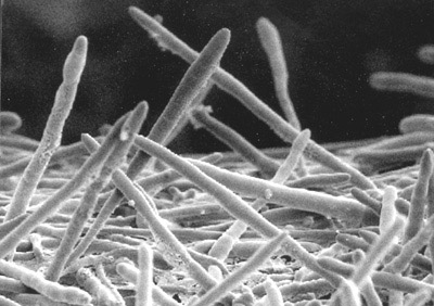
Bacteria are cells without nuclei, therefore they are called Prokaryota.

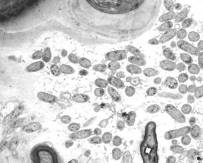
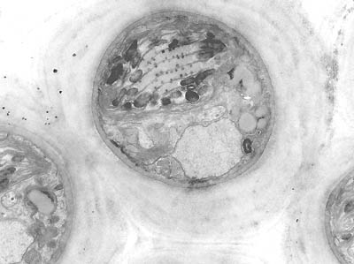
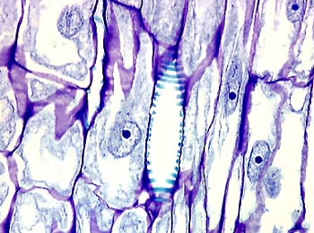
TEM (Transmission Electron Microscopy) image. Magnification 5.000x

A group of oval to elongated bacteria lie between algae (upper part) and two fungal hyphae visible at the lower part of the image. These bacteria have no membrane-bound nucleus. The genetic material inherited is dispersed through the cytoplasm. Bacteria usually form large communities within their niche and replicate very rapidly.
---

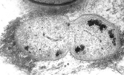
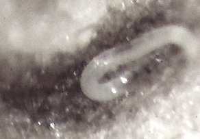
Note the central vertical constriction. Soon we would have two bacteria! A mature and rather old bacterium will rejuvenate in this manner. There is fibrillar material around the bacterium. This is the extracellular matrix (ECM) in which they live. Most bacteria and other cells simply produce their own typical extracellular matrix as an inseparable part of their environment. TEM. Magnification 20.000x
---

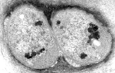
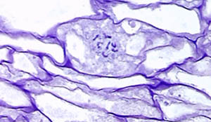
This TEM image illustrates that the division is complete and we have two daughter bacteria from the old one. Note the cell membrane delimiting their cytoplasm from the ECM. Magnification 20.000x
---

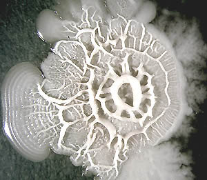
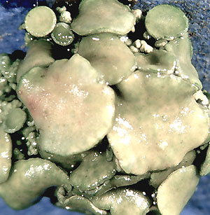
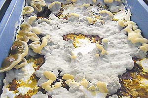
Bacteria can be cultured in Petri dishes containing suitable food stuff.

---

Nucleus-bearing cells are Eukaryotes which may represent a form of symbiosis. There are good reasons to believe that nuclei, mitochondria and possibly some other cell organelles like flagella have a symbiotic origin. Amoeba are unicellular living beings with nuclei, therefore they belong to Eukaryota like all other nucleus-bearing cells do. Algae possess a green pigment, the chlorophyll, enabling them to photosynthesise. They belong to an inhomogenous group of living organisms called the Protista, with both unicellular as well as multicellular members.

---

This is a TEM image of a spherical alga containing a nucleus at the lower part and a chloroplast with grana and thylakoids, a stalk of membranes. Magnification 7.000x
---
An example of a pond water organism belonging to the genus Oedogonium identified by my fellow microscopist Mr. Jan Parmentier who is a well-known expert to most of Micscape readers. (See: Fritsch, The Structure and Reproduction of the Algae. Vol. 1, p. 297.) Note that I put a drop of very diluted solution of Toluidine blue into the sample water! The stain has partly been absorbed in vivo!
---

A colonial life form; Pediastrum boryanum, as identified by J. Parmentier.

---

Fungi are very diverse; you may find unicellular or multicellular fungi, you may also encounter fungi both microscopic as well as macroscopic in size e.g.  mushrooms. Lichens are nice associations not only between algae and fungi, but mostly in the presence of bacteria as well.

---

Just a close-up of a lichen. The green colour arises from the component algae, the cup shape morphology is related to the host Ascomycete fungus.
---

We can, of course, cultivate our edible fungi:

Young, recently developing fruit bodies (Yellow and plate structures sometime with a brown surface, left side) of Ganoderma lucidum cultivated in MES Laboratories* in Horst, The Netherlands.
---

Oyster Mushroom, Pleurotus ostreatus grown in MES Laboratories*

---

Fungi are made of hyphae, the long, cylindrical thread-like cells. They can be grown in Petri dishes and form colonies in a similar way to bacteria. Do you see the threads at the convex-shaped edge of the colony illustrated below?

---

If we experimentally induce a wound in fungal tissues, the hyphae regenerate and try to repair the tissue defect. Repair and regeneration are very essential characteristics of cells/organisms inherited from their ancient predecessors. It ensures the continuity and unity of the cell or tissues. The following image is made using a Low Temperature Scanning Electron Microscope (LTSEM) and shows regenerating hyphae with typical apical tips.

For further details see: Umar, MH & Van Griensven, LJLD (1997). Hyphal regeneration and histogenesis in Agaricus bisporus. Mycological Research 101 (9): 1025-1032.

---

Plants, like algae, are photosynthetic organisms whereas animals are fully dependent to photosynthetic life forms for their existence (this is known as heterotrophism). This very old tree (in Belgium) bears large knots; I am always inclined to designate such irregular swellings as plant tumours!

---

There are so many intermediate forms that we cannot be sure whether a particular member is a plant, animal, algae or fungi. We cannot even classify some of them as uni- or multicellular. They just come together, bound to each other and form colonies. In colonial type of life we can recognize certain fundamental features of tissue forming which we also observe in fungi, plants and animals. A tissue is the cohesive aggregation of the same type of cells. Tissues may come together and form organs or systems which are the main characteristics of fungi, plants and animals. Such cellular variation in form and function can be attained by a phenomenon known as cell differentiation.

---

This is a tissue from the apical part of a young sprout of potato, Solanum tuberosum. Note that the cells are mostly polygonal, cell walls are almost transparent, cytoplasm contains small vacoles and are stained strongly with Crystal violet, an aqueous stain solution of 0.1 %; nuclei are oval to angled and rather lightly stained whereas nucleoli are massive, large and stained dark-blue. This is an example of the young, meristematoid tissue of a plant.

---

This tissue sample is taken from the same potato, but from the central, basal part of the sprout. The cellular components of this tissue are obviously different from the meristematoid top part.

---

You may find there mitotical figures as well.

---

At the more central part of the potato, the cells contain large, round inclusions, a product of great importance i.e. the starch or the amylum. Not all cells are able to make this product. Compare this image with that of meristematoid tissue. These are apparently differentiated cells.
---
Animals are mobile living forms. They have the shortest life history on Earth, but the cellular complexities and developments we find in animals have surpassed all previously achieved accomplishments in any other kind of life forms on Earth.

---

What about this nematode, a worm, Caenorhabditis elegans?

---

A portion of a spleen of a BALB/c mouse, inbred for experimental studies. The dark areas are the lymphoid follicles.
---

In this section of mouse spleen, we see blood forming cells differentiating from less differentiated stem cells. Try to find the mitotically proliferating cells shown here.

---

How different are these liver cells of the same mouse? At the centre there is a vein, the central vena of the lobe. Some liver cells normally possess double nuclei.

---

When stained with PAS (Periodic Acid Schiff Reagent) the cytoplasm show lots of glycogen granulae with magenta-purple colours. At the middle, we recognize the central vein containing erythrocytes; the oxygen carriers of the animal body. A neat example of cell differentiation.

---

There are two resting liver cells on the left and a large, dividing one at the center. Mitosis is in the anaphase. PAS stain.

---

Gall is produced by liver cells and is transported through specific channels to the gall-bladder, in mice or men alike! On the right we see such a biliary duct of the mouse lined by cylindrical, so-called epithelial cells. Around this duct there are cells of smooth muscle and connective tissues, blood capillary at the left, also some lymphocytes, etc. Again, even in one microscopic field as shown here, animal tissues represent a wide variety of differentiated cells.

~~~~~~~~~

Epilogue

We started our tour with prokaryotic cells without nuclei. After a very quick overview we came to the vertebrates, in particular the mammals, to which Homo sapiens sapiens belong. Everybody knows that the diversity of life is enormous. How many species inhabit our world, we do not know exactly! We can only make a rough estimation, several millions of them are surely part of the Earth's biota. Several times more than that number must be gone for ever by extinctions that occurred in the geological past. It's astonishing that all life forms, from the simplest life to man have always been genetically coded in ONE particular cell; whether these are parent cells, bacterial or fungal spores, plant seeds, or fertilized animal eggs. In summary, life on Earth is characterized by two main life forms: unicellular and multicellular. There are still other transitional forms in between these two extremes. By gradual evolution, the pathways that led from single-celled towards multicellular organisms, exhibit an amazing diversity of forms and life styles, and wonderfully illustrates how bio-organization occurs over a very long time span.

Comments to the author M. Halit Umar are welcomed.
 

~~~~~

Some interesting Internet Links:

Cell: http://www.fwkc.com/encyclopedia/low/articles/c/c004001165f.html

Kingdom Protista: http://arnica.csustan.edu/boty1050/Protista/protista.htm

Phycology in Southwest Missouri State University: Southwest Missouri State University, Department of Biology, BIO 530- Phycology

Denis Kunkel's Microscopy: Dennis Kunkel's Microscopy


~~~~~~~~

The author's affiliation:

** MES Laboratories, P.O. Box: 6042,

5960 AA HORST, The Netherlands.

Phone: (0031) 77 464 7575 , FAX: (0031) 77 464 1567

All images presented in this article are registered and copyright to MES Laboratories.

~~~~~~~

Microscopy UK Front Page
Micscape Magazine
Article Library


© Microscopy UK or their contributors.

Published in the September 2000 edition of Micscape Magazine.

Please report any Web problems or offer general comments to the Micscape Editor,
via the contact on current Micscape Index.

Micscape is the on-line monthly magazine of the Microscopy UK web
site at Microscopy-UK

WIDTH=1


© Onview.net Ltd, Microscopy-UK, and all contributors 1995 onwards. All rights reserved. Main site is at www.microscopy-uk.org.uk with full mirror at www.microscopy-uk.net.