The beginner in the study of desmids
may not at first realise that there are desmids that form filaments, some
quite long filaments, and he/she may not always distinguish these from
other algae that is filamentous. The majority of filamentous species of
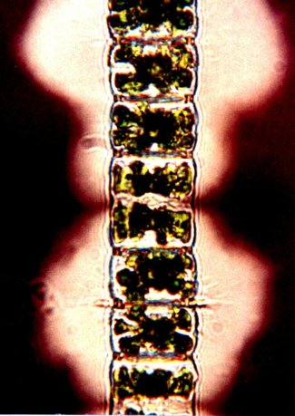
algae have continuous walls divided into cells by septa, for example the
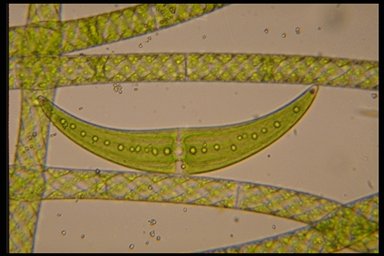
well known Spirogyra which is
related to the desmids, as it also conjugates. (Fig.1 shows Spirogyra
with the desmid Closterium moniliferum).
Examination of the desmid filaments
will reveal that the cells are not contained in a continuous wall but are
separate cells joined at their apices in some way, some tightly with no
visible space between them, others have pads that join the cells leaving
a space, others are loosely joined with interlocking processes. Some genera
exude copious amounts of mucilage that appear to be instrumental in keeping
the desmid cells attached.
The filamentous desmid genera/species
most frequently found are Bambusina brebissonii and Hyalotheca
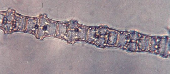
dissiliens. Bambusina brebissonii Kütz (1849) is listed
in early books as Gymnozyga moniliformis Her. (1841). There is only
one species of the genus found in Britain; cells 25-35 µm long 15-23
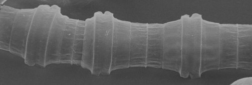
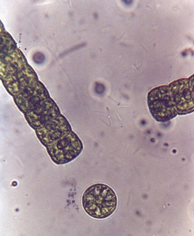
µm broad, circular in apical view. Fig. 2 is a light microscope photomicrograph,
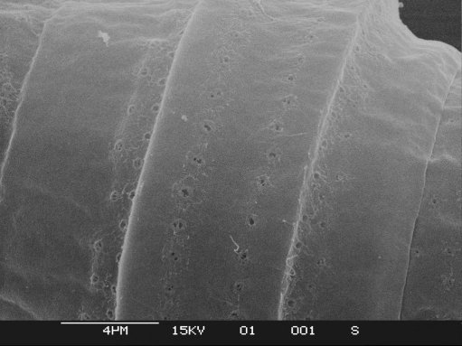
figs. 3 & 4. SEM images. A complete cell is shown between the lines.
In fig. 4. rows of evenly spaced small punctae can be seen.
Hyalotheca Bréb. ex
Ralfs (1848): There are
five British species (Fritsch 1927), H.dissiliens being the most
abundant. (Fig.5 photographed in Indian ink to show the copious mucilage.
Fig. 6 shows one cell broken away and seen in apical view.)
Photomicrographs of other genera/species
of filamentous desmids will be shown later.
The light microscope photos are
by the author, SEM photos are by Andrew Syred.
Reference
A Treatise on the Freshwater Algae
by G.S.West (1904) revised by F.E. Fritsch (1927), Cambridge University
Press.
All comments to the author Bill
Ells are welcomed.
'Desmids that form filaments, part
two' by the author is here.
Images