The little star in the pond by Chitchai Chantangsi, Thailand |
|
|||
When
you look upon the sky by night, you can find the stars, which reflect their
light through the heavens. But do you know that you can also find this
in aquatic dwellings?
In many greenish freshwater habitats, if you collect water samples from there and observe them under a microscope, you may see an amazing microscopic world. By the magnifying power of a microscope, many tiny organisms, such as bacteria, algae (especially green algae), protozoa and other small animals are discovered. |
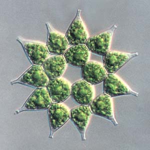
Figure 1>>> Pediastrum simplex; a stellate colony with single process peripheral cells and perforations. Image taken by the Nomarski technique, 40x objective (90 microns in diameter). |
|
Green
algae are predominantly freshwater; only 10% of them are marine, whereas
90% are freshwater (Boney, 1975 and Lee, 1989). They and many other algae
play an important role for all beings; that is oxygen production by a photosynthetic
process.
Pediastrum
is
a genus of green algae that is commonly found in many freshwater microhabitats
because it has a cosmopolitan distribution. Many species of these algae
have been described. They are typically planktonic organisms, drifting
and floating about in ponds, marshes, pools and lakes.
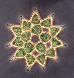
<<<Figure 2. Pediastrum simplex, a fascinating "shining star" when imaged by phase contrast microscopy. 40x objective (90 microns in diameter). |
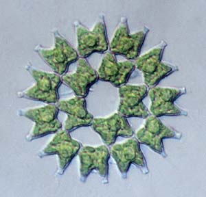
Pediastrum (Gr. pedion, flat or plain + Gr. astron, star) (Brown, 1956 and Bold, 1985) means a plain star. This name implies its shape. The genus is a one-celled thick colonial form, which consists of many cells as many as 2 (rarely), 4, 8, 16, 32, 64, 128 cells or multiple of 2, depending on the species (usually 4 to 64 cells). Most species are a star-shaped colony that is a flat, circular or sometimes irregularly subcircular configuration (Prescott, 1978). A colony comprises two different cell shapes those are single layer or many layers of the inner non-processed cell and single layer of the outermost processed cell. The peripheral cell has one, two or three processes for each species and therefore types and shapes of this cell, including figures and numbers of projections are very important characteristics for species identification. In addition, presence or absence of perforations and ornamentation of the cell wall should be considered for taxonomic contemplation (Smith, 1933). |
Figure 3. Pediastrum duplex, with two-process outer cells and perforations within a starlike coenobe. It was photographed by Nomarski microscopy, 40x objective (65 microns in diameter). |
Asexual
reproduction of this alga occurs by autocolony formation. In this process
the biflagellate zoospore, a motile spore having two flagella, are produced
by the cell in a colony and enveloped by a vesicle that is the emergent
innermost layer of the cell wall (Bold, 1985). Although every cell can
generate zoospores, all cells can not produce them simultaneously. Zoospores
take place by a repeated mitotically nuclear division without cytokinesis,
the cytoplasmic division, and thus the parental cell is multinucleated.
After that, the cytokinesis for every nucleus occurs synchronously and
many uninucleated daughter cells transform into biflagellate zoospores
that swim freely within a vesicle. Subsequently, each zoospore arranges
itself to form a new younger colony by supporting of microtubules.
All of these processes are known as 'autocolony formation'. Sexual reproduction takes place by fusion of biflagellate isogametes, morphologically and physiologically similar gametes having two flagella. Isogamy, union of two isogametes, generates a zygote. |
By
the reproductive process, Pediastrum has produced its descendants
from generation to generation for a long time. Its fossils were discovered
and indicate its existence in many ages, such as Lower Cretaceous (146-113
million years ago), Upper Cretaceous (113-65 mya) and Middle Miocene (14
mya). Amazingly, these little stars have reflected their beauty in aquatic
micro-environments for many millions of years.
Now,
are you ready to investigate them with your own eyes?
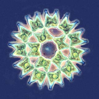
Figure 4.>>> Pediastrum duplex has bright green chloroplasts within each cell for photosynthesis. Phase contrast, 40x objective (65 microns in diameter). |
|
Acknowledgments
I am
grateful to many people. First of all, Wim van Egmond has persuaded and
encouraged my first on-line publication and his kindness for this web page.
To Dr. Chanpen Chanchao, a nice teacher, and Brandon Jones, a good friend,
for their kind advice and improvement of the text. Finally, I am particularly
indebted to Micscape to give me a good chance for this publication.
References: Bold, H. C. 1985. Introduction to the Algae: structure and reproduction. 2nd ed. The United States of America: Prentice-Hall. Boney, A. D. 1975. Phytoplankton. London: Edward Arnold. Brown, R. W. 1956. Composition of Scientific Words. Washington: Smithsonian Institution Press. Hoek, C. van den., D. G. Mann and H. M. Jahns. 1995. Algae: An introduction to phycology. Great Britain: Cambridge University Press. Lee, R. E. 1989. Phycology. 2nd ed. The United States of America: Cambridge University Press. Prescott, G. W. 1978. How to Know the Freshwater algae. 3nd ed. The United States of America: Wm. C. Brown Publishers. Sharma, O. P. 1986. Textbook of Algae. New Delhi: Tata McGraw-Hill Publishing. Smith, G. M. 1933. The Fresh-Water Algae of The United States. The United States of America: McGraw-Hill Book.
|
All comments to the author Chitchai Chantangsi are welcomed.
Please report any Web problems or
offer general comments to the Micscape
Editor,
via the contact on current Micscape
Index.
Micscape is the on-line monthly magazine
of the Microscopy UK web
site at Microscopy-UK