Amoebas are more than just blobs - page 3
SHELLED AMOEBA
by Wim van Egmond, The Netherlands
 

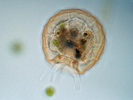
Arcella gibbosa, its test is about 0.1 mm. in diameter.
   
An important group of amoeba make a protective shell called a 'test'. These shelled amoebas can be collected with the same methods as 'naked amoeba' Some caution must be taken when examining them since the tests easily break. I always add some dots of Vaseline under the corners of the cover slip. It often takes some minutes before disturbed shelled amoeba become active

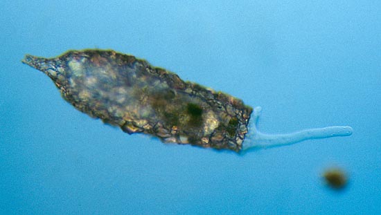
The image shows how the shelled amoeba Arcella is attached to its house by thin pseudopods. Bigger pseudopods are extended through the small opening at the bottom of the test.


Shelled amoeba of the genus Arcella make delicate tests. When observed with a high power objective (40X), the fine texture of the test can be seen. They construct their test by glueing tiny bits of inorganic detritus together.

The bottom image shows the much cruder design of Difflugia, another common shelled amoeba. The sand particles that are used, obscure the view, so the living cell of the amoeba can only be observed when it extrudes its pseudopods.


Difflugia, its test is about 0.3 mm. long.


Page 1: Amoebas are more than just blobs


Page 2: LOCOMOTION AND FEEDING
Page 3: STUDYING TINY AMOEBA
 

All comments to the author Wim van Egmond are welcomed.

Find out more about microscopic pond life on The Smallest Page on the web
The Pond Life Identification Kit may help you identifying small freshwater organisms

Visit Wims home page for links to his many web pages on microscopy

Microscopy UK Front Page
Micscape Magazine
Article Library


WIDTH=1

© Microscopy UK or their contributors.

Published in the September 2001 edition of Micscape Magazine.

Please report any Web problems or offer general comments to the Micscape Editor,
via the contact on current Micscape Index.

Micscape is the on-line monthly magazine of the Microscopy UK web
site at Microscopy-UK


© Onview.net Ltd, Microscopy-UK, and all contributors 1995 onwards. All rights reserved. Main site is at www.microscopy-uk.org.uk with full mirror at www.microscopy-uk.net.