If the resolution of the camera is set to the maximum (640 x 480), and the camera placed over the eyepiece in contact with the microscope, the outer limits of the eyepiece field will be seen, and a centred position can be found which does not vignette the image. How the camera can be held in this position (all sorts of jigs, templates and holders will suggest themselves) can be left to the ingenuity of the operator. The two rubber bands shown in the picture above represent a minimal solution, which only works if the rather stiff USB cable is held captive (BlueTak or tape) as shown. Obtaining still images or video sequences without a USB connection to the computer will of course require an accurate and reliable mounting setup, as no image is available for checking the camera alignment. Image resolutions and video length are also limited to the default values of the camera in stand-alone mode (26 frames at 640 x 480, 107 frames at a heavily pixellated 320 x 240). The simple folding tripod shown enables single-handed manipulation of the specimen, and allows the other hand to access the focusing adjustment on the underside of the instrument. It also provides a convenient place to anchor the USB cable, reducing the risk of damage to, or misalignment of, the camera. If the microscope is used in the same orientation as for
visual use, the software allows for the vertical movement of the specimen
to be reversed, giving a sense of movement familiar to all practised users
of the laboratory microscope. (Reversing the horizontal motion instead
will result in an action more familiar to users of low power stereo microscopes,
which do not invert the image).
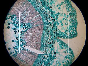
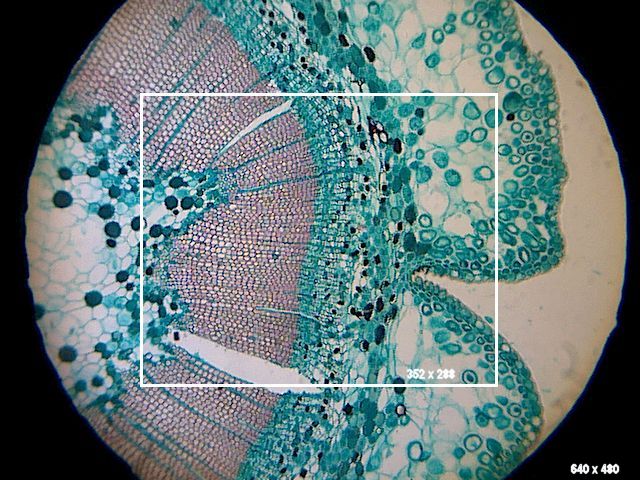
The picture on the right shows the relative field areas seen by the camera, positioned over the eyepiece of an OU microscope, at 640 x 480 resolution (large picture) and 352 x 288 resolution (inset picture), illustrating the points made regarding image size and resolution. The subject is a double-stained cross-section of a yew
stem taken at the lower power of the microscope.
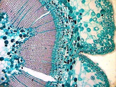
Here is a processed image showing the maximum area obtainable (80KB) from the high-res image. The corners of the picture almost touch the circle of the eyepiece diaphragm. Here is a link to a short movie sequence (1MB) showing the feeding behaviour of the protozoan Paramecium, videoed at a resolution of 352 x 288 under computer control, an image corresponding to the area of the inset in the picture above. The image was shot at the higher power of the OU microscope, and has not been digitally processed except for the reduction to 256 colours necessary to prepare an animated GIF. In conclusion, the results from this equipment are unlikely to win any prizes for photomicrography, but given that they are easily obtained on location with affordable components, they can fulfil the function of a video/still notebook, replacing (to some extent) the need for drawings, and providing images which are conveniently emailable to colleagues for identification and discussion. Microscopy is, more than most, an area in which a picture
is worth a thousand words.
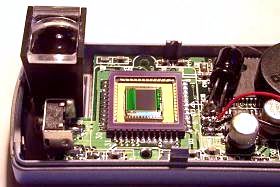
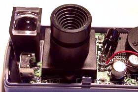
The photographs below are of the camera with the outer casing removed to show the CMOS array with and without the lens assembly. This camera represents better value than many camera modules sold separately to experimenters and hobbyists. Not only is it less expensive than many of these units, unlike them it comes with battery compartment built in, USB cable connection to the computer, and a CD of interfacing software and other useful applications. The CMOS array is sealed with an optical glass window so that it may be cleaned without damage to the array. The lens, unfortunately for experimenters, is glued into its focusing mount.
It can be recommended to anyone wishing to experiment
with odd applications of digital imagery at low cost.
Recently, (late March 2002), Aiptek announced the release of their PenCam Mini 1.3 model, which is a 1.3 megapixel version of the digital camera reviewed here. It is housed in what appears to be the same casing, so presumably has the same desirable physical features of the VGA version, but should be capable of greatly improved image quality. Micrographia will publish a review of the new model when one becomes available. In the meantime, this link will take you to the (Go to Page 1) Contribution by John Walsh, comments welcomed.
Micscape acknowledgement: The author kindly offered this article from his own web site for Micscape to mirror. His own site Micrographia has some splendid resources for the amateur microscopist. Click the author's web site logo below to visit it.
Microscopy UK Front
Page |
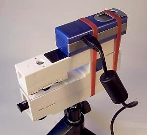 When the camera is connected to a notebook computer, the supplied software
provides a moving image on the computer display allowing accurate positioning
and focusing of the camera.
When the camera is connected to a notebook computer, the supplied software
provides a moving image on the computer display allowing accurate positioning
and focusing of the camera.
 Here are some example shots using the setup described above.
