 |
 |
Flatfield imaging The basics and some personal thoughts By Paul James (uk) |
Anyone who starts observing through the microscope soon realises that the central zone of the field provides the best quality imagery. For eons this was accepted as being a consequence of requiring the ultimate resolution from the optical system, for it was a simple matter to move the subject into the centre, or part of the specimen to utilise the maximum imaging potential of the instrument. As medical progress required more from the instrument, especially in quantitative studies, like scanning histological specimens and examination of smears of all kinds, the need for flatter field designs became the priority for manufacturers. The fact that a flatfield image with all the observable detail apparent without recourse to refocussing and stage manipulation made for a more efficient and more accurate assessment of the image. In short the time involved in observational work could be reduced, and of course fatigue which inevitably leads to mistakes, could be minimised too.
There is nothing new about the desire for flatfield imagery, but it's general incorporation into optical design was accelerated by the medical research's need to satisfy the world's health service's ever growing responsibilities in our society.
Swings and roundabouts
The real problem regarding the design of the perfect flatfield is simply that of the constraints of resolution and field flatness. A camera's lens is designed to provide as flat a field on the film or CCD as possible. It is a simple fact though, that in order to achieve this some loss of ultimate resolution has to be accepted. In camera lenses this is not a significant problem as a highly corrected lens will yield a flat field image which provides enough contrast and resolution for photography. In microscopy the problem is a critical one for resolution is paramount and its reduction in an objective which has been designed to yield a flatter field is compromised.
As with most things in life, money is at the heart of this, and whilst good flatfield achromat imagery can be designed into an optic for a modest cost increase over standard optics, the marriage of high na and flatfield is costly as many more elements are needed in the objective. These are usually more perfectly corrected too viz fluorite and apochromat, and so their complexity accounts for astronomical costs when new. As many as 10 or more elements of glass are used to achieve this state of optical perfection, which translates into 20 or more small optical surfaces which are then held with great precision in the body of the objective.
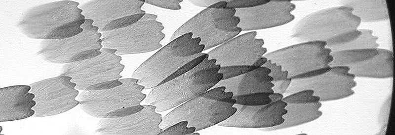
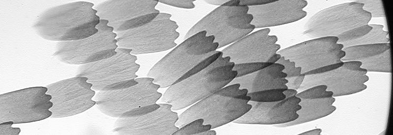
Comparison of field with x 10 non Plan and x 10 Plan objectives
NB The results from the digital camera show less field curvature with non Plan objectives than observed with the eye through either the same eyepiece or through the trinocular light path. Whilst this is a very welcome 'flaw' in my setup it should be noted that this is not necessarily a universal feature of all camera/microscope combinations.
|
|
The two images above illustrate the basic difference between Plan and non Plan objectives. The Plan form at bottom maintains focus on butterfly scales to the edge of the field. The na of the x 10 non Plan is 0.40 and the x 10 Plan is 0.25 and this is very obvious in visual use. Note that the quality of the non plan image is very good at the field edge too when focussed for this zone.
Eyepieces and Plan objectives
The Plan objective's role is to project a flatfield image to the eyepiece, and when the manufacturer's wide field eyepieces are used in conjunction with their range of Plan objectives, the results can be stunning. Most other eyepieces will invariably provide a flattish field too, but cannot be expected to compare with a matched combination. Some microscopes have inbuilt field flattening optics just above the objective changer which can influence the final result when using other manufacturer's lenses. So this must be taken into consideration if anyone wants to radically change to Plan objectives on an instrument so equipped.
Parfocality
In the pursuit of optical excellence the amateur can easily forget temporarily at least, the important issue of parfocality. One may collect a turret full of very good optical hardware over time from different manufacturers and eras, but the result can be a hotpotch with the very real problem of causing damage to either slide or objective..... It only takes one mistake to cause some damage. Having experienced many years of the necessity of treating individually every objective which was not parfocal on the turrets of my first microscopes, I know only too well the time wasted when revolving objectives into position with great concern regarding it focussing. Today parfocality is accepted without question by most novices. Having this simple yet useful facility often reminds me of those past earlier years.
In a nutshell it might seem prudent to obtain a couple of Plan objectives initially, but if they are not parfocal on your stand then the pleasure of their use might be diminished by the constant care needed to avoid damage when revolving the turret ?
The Dilemma
In the natural run of events for those thinking of tweaking their 'scopes to suit their purposes, it follows that there are several constraints which impose themselves as soon as we start to make decisions.
My personal rationale and setup
I can only cite my own philosophy here regarding decisions that have led me to employ the use of flatfield objectives. My reasoning is as follows :-
The problem in using achromatic objectives, flatfield or otherwise is that the x20 upwards require the use of compensating eyepieces for best results, and those optics below the x 20 require Huygen type eyepiece forms.
Achromatic imagery especially in low to medium power is more than adequate for my purposes BUT the need to change eyepieces for higher power is irksome and results in dust finding its way into the eyepiece/binohead as well and offers the remote but real possibility of damage caused by accidental knocks etc..
The way round this problem for me was to acquire fluorite objectives throughout the range so that compensating eyepieces would not only yield corrected imagery for all powers, but that they can be left permanently in situ. I wanted flatfield imagery for low to medium power, but for higher powers such as the x40 and x50-100 standard fluorites would suffice, since these optics were to reveal detail and not large flat sections across the field. Rarely if at all do I come across specimens that fit the field at high power which are truly flat and level anyway, so it seemed to me that my money would be more effectively spent by getting flatfield for low power work and standard fluorites higher up the range. So my arsenal of optics of PLAN Fluotars x3,x6,x10,x20 and STANDARD Fluotars x40 and x50 oil form my objective battery. The complete set took quite a time to accumulate, but I am very satisfied with the overall optical quality and in my case the upper limiting aperture of about 1.0 na which dry condensers can cope with.
I have deliberately omitted any comments concerning the costly Plan Apochromat objectives of high numerical aperture as they are invariably very expensive and their excellent properties are only marginally greater than the fluorite groups.
Conclusion
The slight reduction in resolution of the Plan fluorites compared to the standard fluorite range I find of little consequence in general usage with the x 10 ocular, and any shortcomings are not noticed in dynamic observations of living organisms. Indeed the proper utilisation of the higher na image cannot be achieved with less than x15 eyepieces, and whilst this combination on static preparations is satisfactory, the marginalisation of depth of field with higher na can be an impediment....the "catch 22" in observing dynamic subject matter.
Justification of expenditure on flatfield objectives and eyepieces is entirely a personal matter. I have been able to happily rationalise all this for my own purposes. Your personal requirements may differ, but I hope these short notes have clarified some of the issues concerned. with flatfield imaging.
Ultimately all this has increased my observing pleasure, and this is of course one of my prime goals.
| All comments welcome by the author Paul James |
Microscopy
UK Front Page
Micscape
Magazine
Article
Library
Please report any Web problems or offer general comments to the Micscape Editor.
Micscape is the on-line monthly magazine of the Microscopy
UK web
site at Microscopy-UK