|
|
A Gallery of Maleic Acid Photomicrographs (using a variety of illumination
techniques) |
|
|
A Gallery of Maleic Acid Photomicrographs (using a variety of illumination
techniques) |
Maleic
acid is an industrial chemical used mainly in the production of
synthetic resins, and as an intermediate in the production of other
chemicals. It is sometimes called Toxilic acid, probably due to the
unpleasant results of exposure. The MSDS safety data for
this compound compose a litany of nasty consequences for the unwary
user. It is harmful if ingested, inhaled or absorbed through the
skin. The chemical readily destroys tissues of the mucous
membranes, skin and eyes.
As can be seen in the structural
formula below, maleic acid contains two COOH carboxylic acid groups,
and is therefore referred to as a dibasic
or diprotic acid.
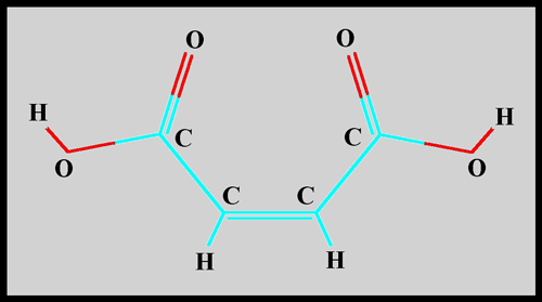
The following image shows the
molecular shape.

Since the melting temperature of
the white crystalline solid is quite low, about 135 degrees Celsius, it
is possible to prepare a melt specimen by gently heating a very small
quantity between slide and cover-glass. Note that I do not recommend doing so!
(See above.) My slides were prepared in the lab using a fume hood.
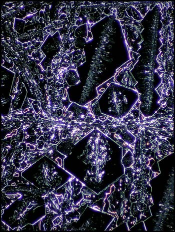
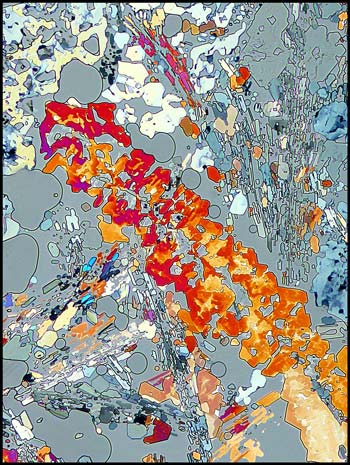
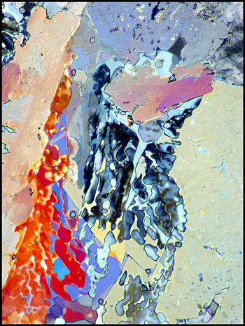
The first image in the article
gives a good idea of what a typical maleic acid field looks like.
Many crystal structures are surrounded by a rather random matrix of
amorphous material. Under dark-ground illumination, the two types
of material can be seen clearly.
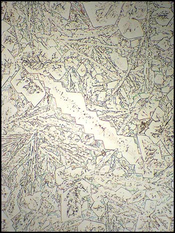
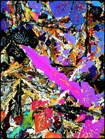
An ordinary transmitted light image
of a field is shown below on the left, and to its right, the identical
field between crossed polars.
If the melt is photographed during
the process of crystallization, one can see the tell-tale lines between
liquid and the bubbles that invariably occur.
The image on the left uses
polarized light, (with crossed polars), while that to the right uses in
addition, two lambda/4 compensators in order to produce the white
background.
If a
small section of the field below is observed under much higher
magnification, interesting detail can be seen. (bottom two images)
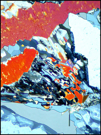
The
unusual field below left was photographed between crossed polars, with
one lambda/4 compensator. That on the right used, in addition, a
lambda compensator.
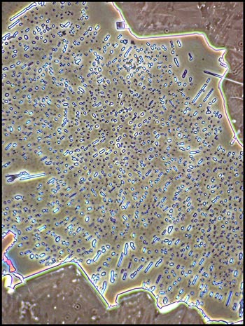
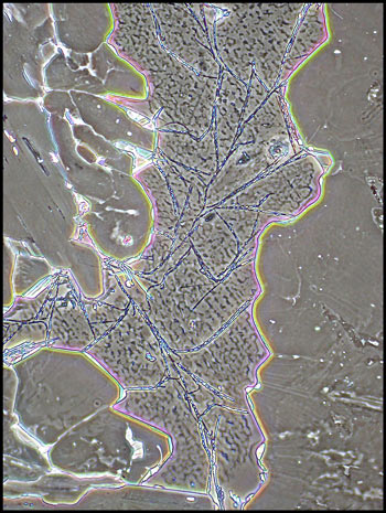
Although phase-contrast
illumination is usually used to view biological specimens, it can
sometimes provide an interesting perspective on the hidden detail in
crystal structures. Note that the three images below are at a
higher magnification than the other examples in the article.
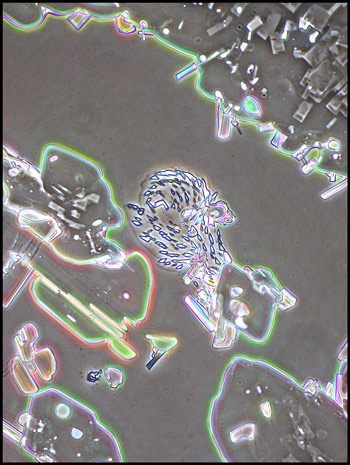
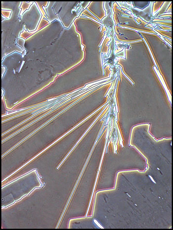
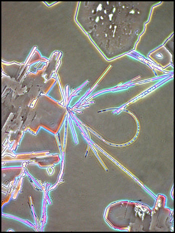
I noticed in a couple of the
prepared slides, that strange, very thin straight and curved structures
had formed. These can be seen in the image that follows.
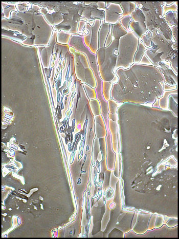
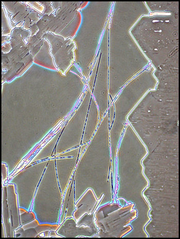
When these structures are viewed at
higher magnification, using phase-contrast illumination, striking
details are resolved.
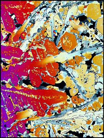
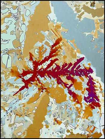
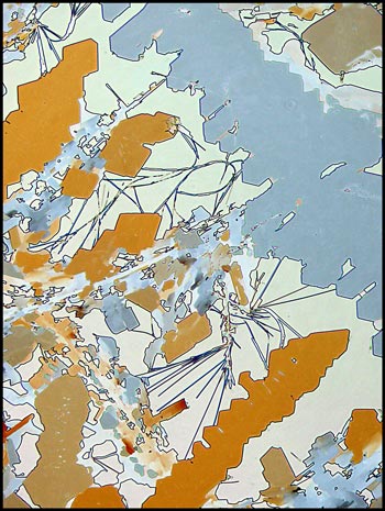
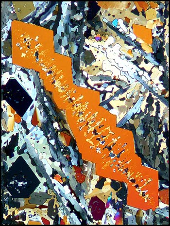
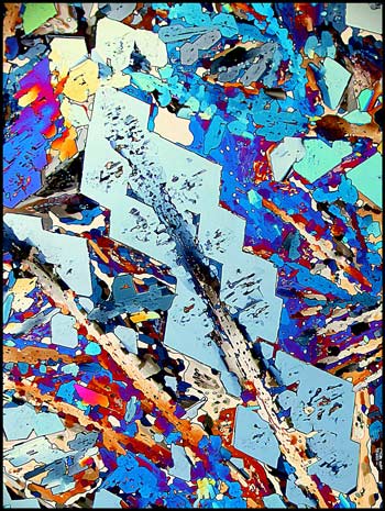
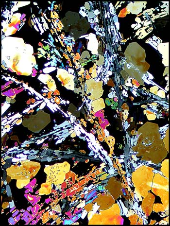
Many of the large crystal
structures that form seem to be based upon a series of imperfect
diamond shapes that have grown together. Compensators were used
to change the colouring of the two images.
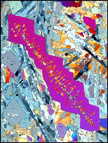
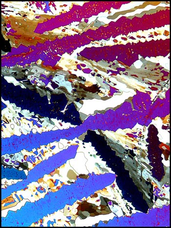
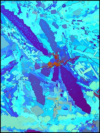
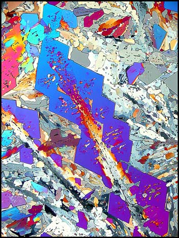
A similar example is shown below,
again with the use of compensators to alter the appearance.
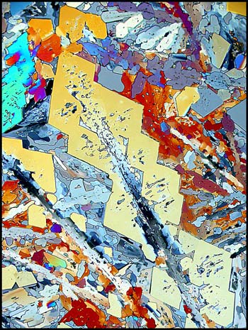
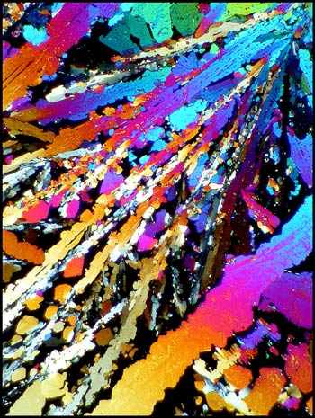
The final image shows the
characteristic that I most dislike about maleic acid melt crystal
structures. Randomness! The fields also tend to be filled
with unsightly garbage, and this makes finding artistic images
difficult!
Photomicrographic
Equipment
The images in the article were
photographed using a Nikon Coolpix 4500 camera attached to a Leitz
SM-Pol polarizing microscope. Images were produced using several
illumination techniques: transmitted light, dark-ground
illumination, phase contrast and polarized light. Crossed polars
were used in all polarized light images. Compensators, ( lambda
and lambda/4 plates ), were utilized to alter the appearance in some
cases. A 2.5x, 6.3x, 16x or 25x flat-field objective formed the
original image and a 10x Periplan eyepiece projected the image to the
camera lens.
Published in the
September
2005 edition of Micscape.
Please report any Web problems or
offer general comments to the Micscape
Editor.
Micscape is the on-line monthly magazine
of the Microscopy UK web
site at Microscopy-UK
© Onview.net Ltd, Microscopy-UK, and all contributors 1995 onwards. All rights reserved. Main site is at www.microscopy-uk.org.uk with full mirror at www.microscopy-uk.net .
