|
White On Black: Reverse Ink Drawings by Richard L. Howey, Wyoming, USA |
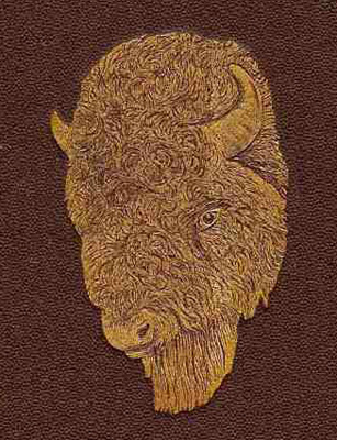
Recently I acquired a couple of volumes of The American Naturalist: A Popular Illustrated Magazine of Natural History. The covers were detached, the spines were missing, some pages were torn around the edges, and others were so loose they could be pulled out with the merest tug. In this essay, I am going to discuss Volume 1, published by the Essex Institute in Salem Massachusetts in 1867 and 1868. You might well wonder why anyone would bother with such a scruffy old volume. The cover is a rich dark brown with a marvelous drawing of a bison head in a shimmering gold color.
Then, consider a few of the topics in the table of contents: (the number is simply for convenience and has no relation to the text)
1) The Land Snails of New England
2) The Fossil Reptiles of New Jersey
3) The Moss-Animals or Fresh Water Polyzoa
4) Some Errors Regarding the Habits of Our Birds
5) The Food of Sea Urchins
6) The Tarantula Killers of Texas
7) Parasitic Plants
8) The Habit of the Gorilla
9) The Sea Horse and Its Young
10) Something About Jelly-Fishes
11) The Hand as an Unruly Member
12) Desmids and Diatoms
13) The Southern Muscadine Grape
14) Notes of a Fur Hunter
15) A Botanical Excursion in my Office
and there are 32 other articles in this volume! Now how could anyone resist such a treasure trove like this–and for only a few dollars at that–for 698 pages which wonderfully includes a glossary telling us that Kjoekkenmoeddings (from an article on shell-heaps) comes from the Danish meaning “kitchen-refuse” and even tells how to pronounce it. There is a certain irony in the fact that even though we now have all of our computer word processing and database technology, almost never do scientific authors provide us with glossaries anymore. Now, to read even a semi-technical biological article, I have to drag out my dictionary of biology and even then I sometimes don’t find the terminology I need and even though I have studied some languages, I am not infrequently at a loss to know how to pronounce certain biological vocabulary.
I should also mention that each issue contains Reviews; Natural History Miscellany, under the four topical headings Botany, Zoology, Geology, and Microscopy consisting of wonderful little notes and observations; a section of correspondence which allows subscribers to contribute their observations on such things as “Preparation of Snail’s Tongue for the Microscope,” “Works on American Lichens,” “Comb-like formations on Birds,” and even one on “Snake Charming.” Also each issue has a section on the Proceedings of Scientific Societies. In addition, there is a list of 16 plates and a second list of 161 woodcuts. From a book collector’s viewpoint, this volume is a disaster–no spine, detached covers, printed on paper that flakes apart, and loose and torn pages. I took some duct tape and “fixed” the binding so that, at least, it now holds together as a book. It’s certainly not attractive, but the contents are and especially the woodcuts and even more especially the plates a number of which were done using the old classic technique of a surface inked black with ink being scraped away to produce the images–thus white drawings on a black background, a very effective, almost dramatic, technique.
In earlier times, naturalists either learned how to draw or found someone who could in order to illustrate their observations. Some of the drawings have become classics and you can see examples of Hudson and P. H. Gosse in Ian Walker’s recent article. Color lithography was a tricky and expensive procedure and many older plates were hand colored. Two remarkable examples of enormously elaborate and intricate drawings are those of Jan Swammerdam and Ernst Haeckel. Swammerdam lived from 1637 to 1680 and his major work The Bible of Nature is most famous for the extraordinary drawings of his detailed and delicate dissections of insects. Some have described him as perhaps the greatest micro-dissectionist who ever lived. It would be a great service if a publisher, such as Dover Press, were to put out a selection of Swammerdam’s drawings as they did for Haeckel.
Haeckel was a man of broad-ranging curiosity and talents. Many specimens which he collected are still to be found in a museum in Jena. He was the great defender of Darwin’s theory of evolution in Germany and wrote extensively on biological evolution. His drawings for Kunstformen der Natur (Art Work in Nature) run the gamut from radiolaria to siphonophores to orchids to tunicates to mammals. Some of the drawings were done from material collected by the Challenger Expedition which is credited for the discovery of 4,500 species of radiolaria alone! The drawings of radiolaria capture their elegant, sculptural forms and reveal to us a stunning set of examples of complex symmetries. Sometimes Haeckel’s drawings have been criticized as being idealized, too perfect, too “artistic”. I must admit that I find them most pleasing and am a great fan of his work. If you would like to see some remarkable high resolution scans, you can find them at this site: http://caliban.mpiz-koeln.mpg.de/~stueber/haeckel/kunstformen/natur.html
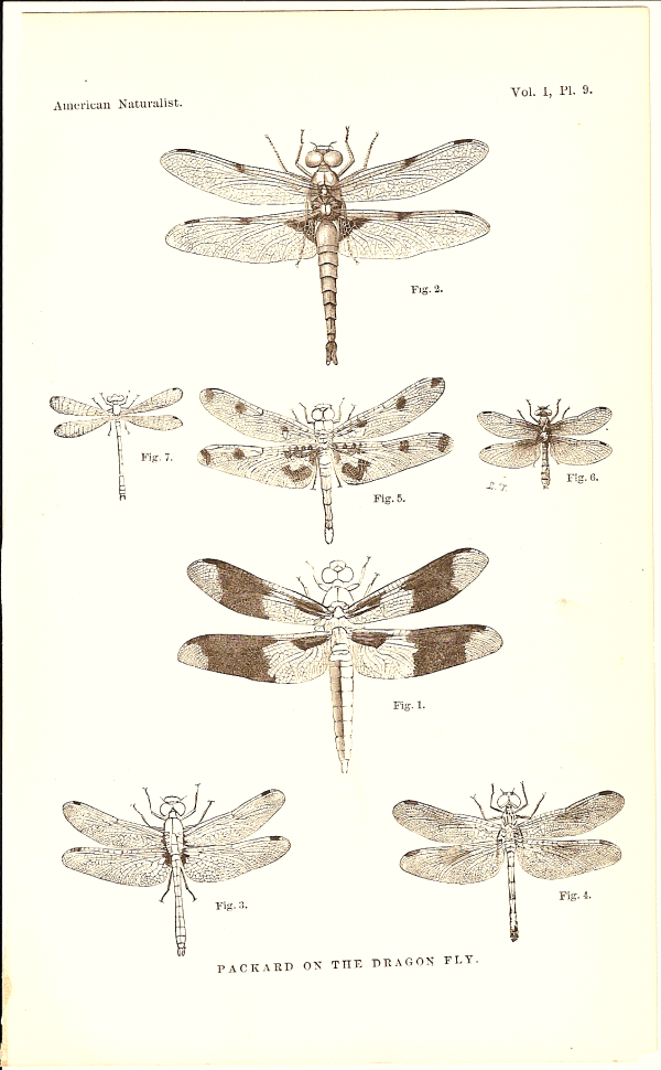
Nothing in my modest volume compares to the Haeckel drawings, but the care and craftsmanship are evident in both the woodcuts and the plates. First let us consider a few of the woodcuts. The first plate is of dragonflies. Whenever I go out to the ponds, lakes, or rivers to collect water samples, I always delight in watching the dragonflies, particularly the way they hover and dart. The large brown species and the large blue species are especially pleasing. This plate has drawings of seven specimens and they are remarkably detailed.
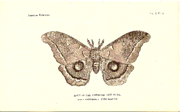
The second woodcut I would have you consider is a remarkable drawing of a male American silk worm moth (Telea polyphemus).
The actual moth is a gorgeous creature, but even from the drawing we can get distinct hints of its beauty and the complexity of its patterning. These are revealed in the distinctive “eyespots” on the hind wings and smaller ones on the forewings. In addition, the bordering near the edges of the wings is striking. The form of the antennae, quite different from most butterflies, is clearly represented. When one looks closely at the body, the fuzzy character derived from many tiny hairs, is clearly portrayed. The bottom edges of the wings are also covered with small hairs and the upper surfaces with minute scales which the drawing also suggests. Overall, a remarkable image.
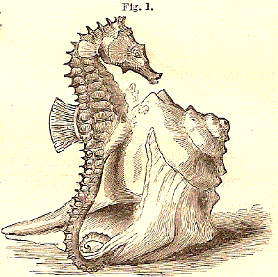
The third woodcut is a small and rather idealized one. It shows a seahorse next to a conch shell. Seahorses are, which we would not expect from their appearance, fish. Their appearance is so charming that many species have been over-collected to the point of endangerment. They are extensively used in kitschy craft items, they are thought by some cultures to have magical and medicinal properties, and are often displayed (and frequently later discarded) just for their unusual aesthetic character. The attribution of “magical” properties is, in large part, a consequence of the fact that it is the male which carries the young in a brood pouch and cares for them until the eggs hatch. Since even small species can produce over a dozen eggs at once, they have come to be regarded as symbols of fertility.
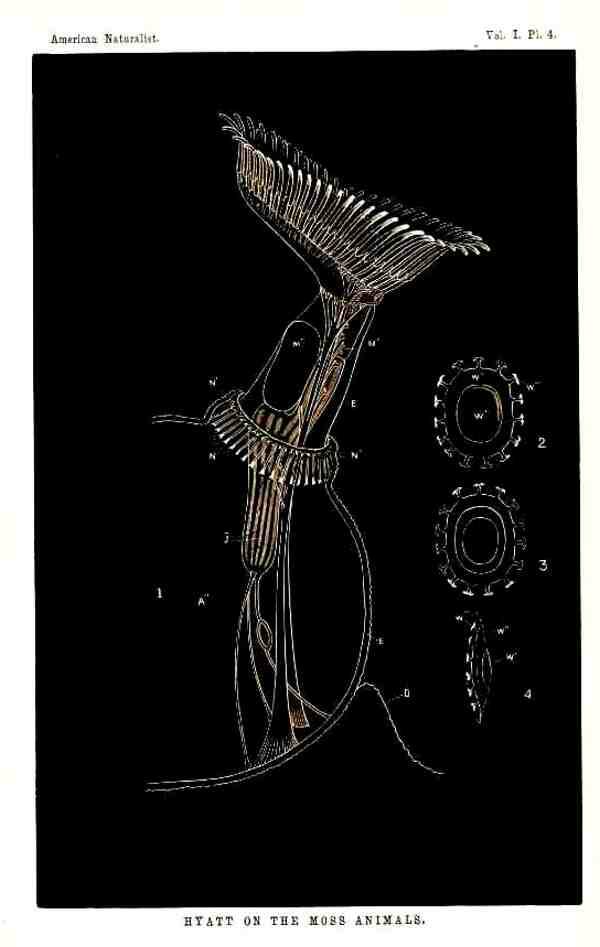
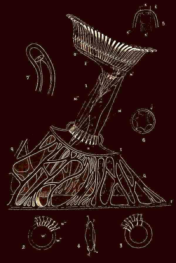
Next, let’s take a look at some of the “scratch” ink plates. The first three plates accompany an article by Alpheus Hyatt on freshwater “Moss Animals” or bryozoa. The organism here described and drawn is Fredericella regina. The first of the three plates has eight figures, the first three of which show colonies of the organism on pieces of bark. I have not found this particular genus, but rather the genus Plumatella repens which was also growing on bark–on the underside of logs floating in a beaver pond. Figure 4 shows the morphology of a complete zooid and figure 5 is a closeup of the tentacles and mouth area. Figures 6,7,and 8 show further details of the mouth area from different perspectives.
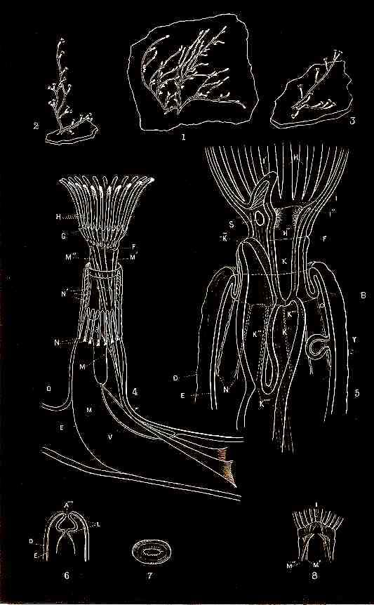
Bryozoa are predominately marine and show a great deal of variation. Although there are relatively few freshwater species, they are among the most elegant and intriguing of freshwater organisms. In the second plate, figure 1 is again a drawing of a single zooid, but from a somewhat different perspective with the tentacles fully extended. Figures 2,3, and 4 show three views of a statoblast of Fredericella. Statoblasts are sort of a cross between eggs and cysts. Statoblasts are remarkably hardy; they can resist both drying and freezing. Sometimes they are abundant along a shoreline and birds provide transportation for them from one lake to another. The statoblasts of Plumatella are not as ornate as those of Fredericella, but are nonetheless distinctive and they can be an aid in identification. Finding statoblasts in a particular pond is, however, no guarantee of finding the colonies. Repeatedly I have waded out into beaver ponds, rolling over log after log, often finding marvelous algal growths, but no bryozoa in spite of an abundance of statoblasts. So, one autumn after a frustrating bryozoanless summer, I decided to try my hand at inducing some statoblasts to “hatch” (and, no, I didn’t sit on them). I placed a dozen or so statoblasts in a standard-sized Petri dish to which I added a standard salt medium (Giese’s salts) or you can use some boiled pond water or artesian water. Just don’t use distilled water, since the lack of salts proves toxic to a number of micro-organisms. Then I added a couple of pipets full of small flagellates and ciliates from rich cultures. Imagine my excitement when several days later I discovered that two of the statoblasts were lying on the bottom of the dish and had thickened so that they looked like an oval-shaped sandwich made of brown bread and egg white. Thereafter I inspected the dish every day and had the pleasure of watching the development of two colonies that attained lengths of between 2 and 3 inches and lasted for several months. This was possible only because I fed them every day from a series of Colpidium cultures which I maintained just for that purpose. It was always a delight to watch the crowns of elegant white tentacles emerge from their tubes and begin to feed. The tentacles are covered with thousands of cilia which create currents steering potential food toward their mouths.
The third plate also shows an individual zooid, but in even greater detail and also has additional figures of statoblasts.
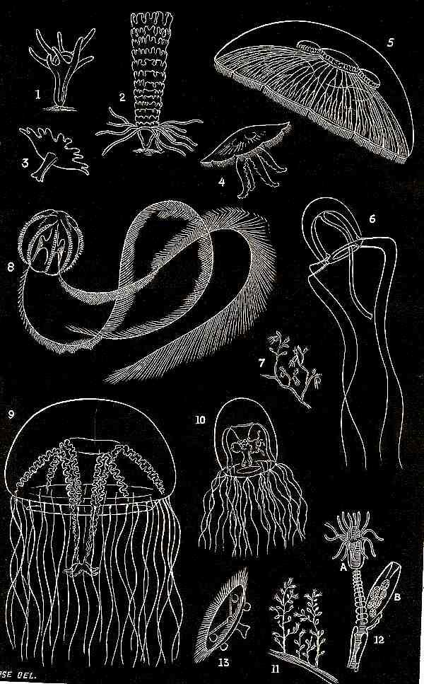
This volume also contains an article by Edward S. Morse titled “Something About Jelly-Fishes”. The plate which accompanies it is quite wonderful and contains 13 figures showing hydroids in various states of development, medusae, and a ctenophore. Surely to late 19th and early 20th Century audiences, these drawings were both delightful and informative.
One of my favorite plates is that for the article by Professor L.W. Bailey “On Desmids and Diatoms”. Even though some of the figures are just bare outlines, they are nonetheless distinctive and a significant aid to classification. Other figures show considerable detail and are quite pleasing to contemplate. I have found these drawings helpful taxonomically and so I am including the list.
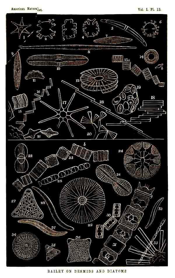
“Explanation of Plate 13
Figs. 1-5 DESMIDS–1 and 3, Euastrum; 2 and 6, Xanthidium, resembling fossils found in flint; 4. Micrasterias; 5 Closterium.
Figs 7-21. DIATOMS–Fresh-water. 7, Navicula; 8 Nitschia; 9, Stauroneis; 10, Pinnularia; 11 Eunotia triodon; 12, Meridion vernale; 13, Tabellaria flocculose, –a, front view, b, side view; 14, Cyclotella kuetzingiana, –b, side view; 15 Acnanthes; 16 Diatoma flocculosum; 17 Astrionella; 18 Bacillaria paradoxa; 19, Mastogloia; 20, Licmophora; 21 Odontidium.
Figs. 22-36. DIATOMS,–Marine Forms. 22, Amphiprora; 23, Amphitetras, forming a zigzag chain,–a, a frustule about to divide into two, b, two frustules newly formed but not yet separated, the “connecting membrane” having fallen off; 24, Asteromphalus, a beautiful deep-sea form, taken from below 2,000 fathoms in the sea of Kamschatka; 25, Podosira; 26 Navicula didyma; 27, Triceratium; 28, Nitschia; 29, Arachnodiscus; 30, Grammatophora; 31, Biddulphia, –a, two frustules still enclosed by the “connecting membranes,” b, “connecting membrane” widening previous to self-division; 32, Pleurosigma; 33, Synedra; 34 Coscinodiscus; 35, Triceratium; 36, Amphitetras.
The forms are not accurately drawn to scale, but are for the most part magnified about four hundred diameters.”
That is a considerable amount of information to be packed into a single plate.
Another article with a striking and informative plate is “The Home of the Bees” by A.S. Packard, Jr., M.D. It opens with a sentence that one would never find in a natural history journal of today: “The history of the Honey-bee, of its wonderful instincts, its elaborate cells and complex economy, have engrossed the attention of the best observers, even from the time of Virgil, who sang of the Ligurian bee.” The plate is of special interest because it provides information about a Humble-bee, a Carpenter bee, a Leaf-Cutter bee, a little green upholsterer bee, and a common green Mason bee, so again I’ll provide the list describing the figures.
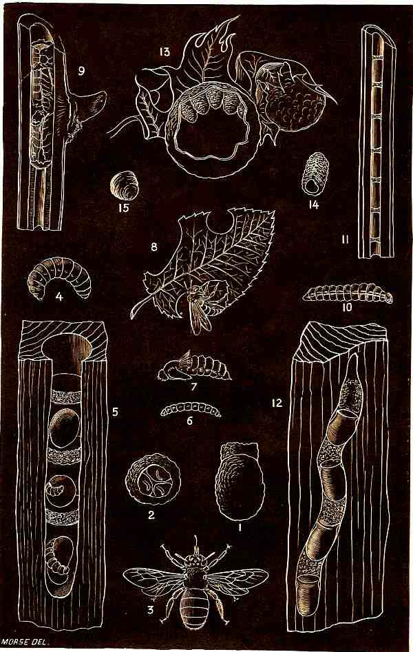
“EXPLANATION OF PLATE 10.
Fig. 1. A cell of the Humble-bee; natural size, with the pollen mass built upon the top.
Fig. 2. End view of the same cell, showing the three eggs laid in three divisions of the cavity.
Fig. 3. Xylocopa Virginica, the Carpenter Bee.
Fig. 4. The larva of Xylocopa Virginica, the Carpenter Bee; natural size.
Fig. 5. The nest containing the cells of the same, with the partitions and pollen masses, on which the young larva is seen in the act of feeding; natural size.
Fig. 6. Young larva of Anthrax sinuosa; side view.
Fig. 7. Pupa of Anthrax sinuosa; side view; natural size.
Fig. 8. The Leaf-cutter Bee (Megachile), on a rose-leaf, in the act of cutting out a circular piece.
Fig. 9. Cell of Megachile, in the elder; natural size.
Fig. 10. Larva of Ceratina dupla, the little green upholsterer Bee; enlarged.
Fig. 11. Cells of the same in the stem of the elder; natural size.
Fig. 12. Cell of Osmia lignivora, new species, the wood-devouring Mason-bee, excavated in the maple; natural size.
Fig. 13. Cells of Osmia simillima, the common green Mason-bee, built in the deserted gall of the Oak-gall Fly.
Fig. 14. A single earthen cell of the same; natural size.
Fig. 15. Pollen mass, or bee-bread of Osmia lignaria; natural size. It is made up of distinct pellets of pollen, which are probably stuck together with saliva.”
The final plate which I’ll include here is for a nine part article on “The Land Snail of New England” by E.S. Morse. With this figure, I shall concentrate only on figures 3 through 7 and provide you with a list of those. For the rest, you can dig out an old textbook or use your imagination. For example, figure 13 is an illustration of a snail bungee jumping.
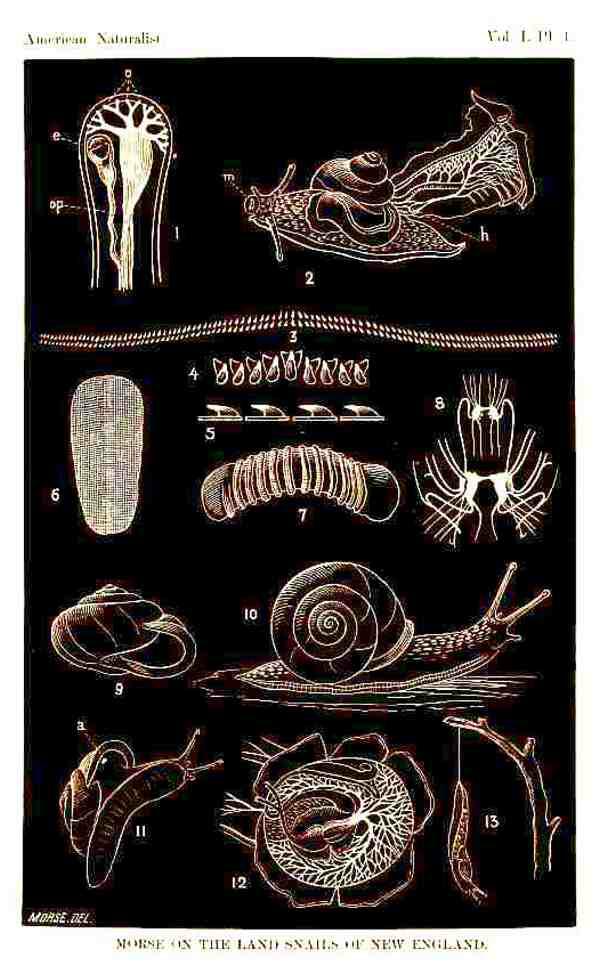
Figures 3-7 are of particular interest to me, since they represent the jaws, mouth parts, “tongue,” and teeth of a snail. The bands of teeth (or radulae) are moved back and forth by an elaborate musculature which scrapes food from a surface.
Any of you who have kept aquaria with snails in them have likely observed thin tracks up the sides through the algal growth. The snails use their radulae to scrape off the algae for food. These snail tongues are marvelous structures; they are calcareous, strong, and very efficient. They make splendid objects to examine under the microscope both with brightfield and polarized illumination. To prepare them, however, you have to have the ruthlessness of Robespierre. The first step is to guillotine the snail. The head is then soaked in a solution of household bleach until the radula dissolves. [Caution: Bleach is a strong caustic, contains chlorine, and is especially dangerous to the eyes and mucous membranes.] This may take several days and fresh solutions of bleach. As is almost always the case when trying to isolate calcareous structures from surrounding tissue, it is best to trim away as much excess tissue as possible with a sharp scalpel before treating it with bleach. After adequate soaking, you will be able to dissect out the radula with little difficulty. In some cases, the radula will be a long thin band up to 2/3 the body length of the snail. This means that you can often cut it into several pieces and make 3 or 4 slides from one radula. You can let the pieces dry and then mount them in balsam or a synthetic resin. [NOTE: You cannot, however, use any lacto-phenol type mountants, since they contain acids that will dissolve the structures you wish to study.]
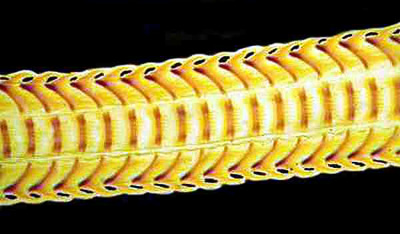
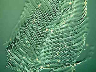
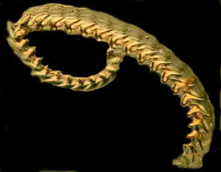
If you get interested in studying radulae, you are by no means limited to land snails. You will find them in aquatic snails–both freshwater and marine, in chitons, limpets, sea-hares, whelks, nudibranchs, etc. Here I’ll include a few of my own images of radulae.
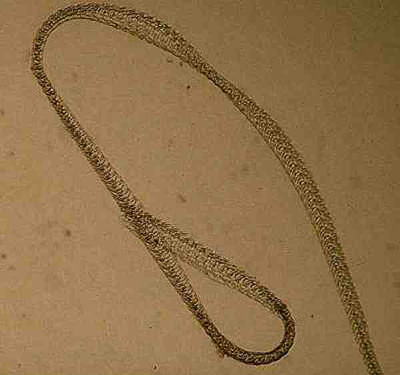
Complete snail radula
Closeup of a snail radula
Radula of a chiton
Radula of a nudibranch
The next time you are in a bookshop browsing the natural history section, keep an eye out for old beat-up copies of early journals. You may find some surprising bargains.
Once again it is time to thank my devoted wife for putting up with my microscopic idiosyncracies for over 45 years now and especially for her help in editing and proofreading my eccentric articles.
All comments to the author Richard Howey are welcomed.
Microscopy
UK Front Page
Micscape
Magazine
Article
Library
Please report any Web problems or offer general comments to the Micscape Editor.
Micscape is the on-line monthly magazine of the Microscopy UK website at Microscopy-UK