 |
| The Fly In The Ointment An Achilles Heel in Köhler illumination? By Paul James |
| A personal exploration into the most commonly installed microscope illumination system |
If you had to choose one aspect of microscopical technique, which at least on the surface many practitioners of the art take as gospel or for granted, then it must surely be the belief in the effectiveness of current illumination techniques such as Köhler's, which has in one way or another become the most commonly installed illumination system. But there are weaknesses in many Köhler's or Critical illumination units for that matter, which have everything to do with the execution of theory into practice, as well as modern trends in making illumination modules compact. Indeed Köhler's theory is flawless as it stands: a high intensity point source expanded to fill the field of view with evenly distributed light. Yet the incentive to improve the uniformity of lighting systems in microscopy in the late 1800's was ultimately a photographic one, since film is far more sensitive to variations of light intensity across an unevenly light field than the eye. That is still as true today with modern films and ccd's as it was in the 1890's when August Köhler contrived his system.
Of course you might not have any problems with unevenly lit fields especially at low to middle powers simply because your lighting system will in all probability be a modified one. If on the other hand you find the filament still vaguely imaged in the background regardless of optimisation when using medium to lower powers such as around x100 downwards, then clearly something is not as it should be.
In order to understand where the illuminating system can fail to provide a truly evenly lit field, we need to look for the weak spots in the system as a whole starting at the filament and through to the specimen :-
|
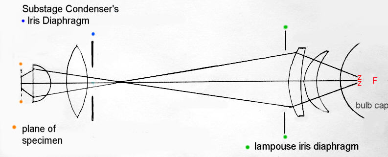
If the microscope's field of view is to be uniformly illuminated by the magnified spot from the white hot filament F then it stands to reason that the optics involved must be reasonably free of aberrations: more especially the spherical form. The above diagram is a simplification of Köhler lighting showing the basic light path from the filament to the specimen plane.
Köhler Illumination is a two stage process: firstly the lamphouse condenser projects an image of the filament very near to the anterior focal plane of the substage condenser, usually where the iris is situated, which then passes through the optics of the substage condenser to form the final image of the source onto the specimen. If this two stage operation is performed correctly then the field of view of a medium power x 10 objective should be filled uniformly with light, in which the specimen is imaged of course. The average size of the field 'disc' projected by the condenser onto the specimen plane is usually 2-4mm depending on the focal lengths of both the lamphouse and substage condensers used as well as the relative distances between the two. Low power observation requires much greater field coverage than this, which can be partially realised by removing the top of the substage condenser thereby extending its focal length.
What is meant by the 'Source'
This is the pivotal point when understanding Köhler or critical illumination. Strictly speaking the word source is used to denote the particular plane in the illumination's optical train which is finally imaged together and coincident with the specimen on the surface of the slide.
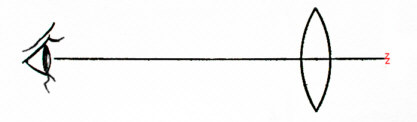
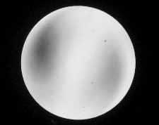
The simplified illustration below shows how the source is perceived by the eye. Here for demonstration purposes the simple lens has been placed near its focal length in front of the filament. The observer will see the whole face of the lens filled with light : This is what we see in the microscope's field of view together with the specimen and is known as the source. However in reality the use of simple single lenses for both lamphouse condenser and substage condenser would result in a very distorted or unevenly lit field or source because of gross spherical aberrations. The second diagram below shows an example of such a distorted source.
 |
 |
When Köhler is setup, the filament distance to the lamphouse condenser is so adjusted that a small area of the filament literally fills the whole area of the lamphouse condenser, and as seen through the microscope's eyepiece will also have the lamphouse iris in focus in the field. So closing this iris will simply mask the field, and not effect the light cone's effective numerical aperture from the substage condenser, but is necessarily effective in suppressing any rogue lighting which can reduce specimen imaging contrast. The source is therefore the area of light in the zone encompassed by the field iris. In fact the plane of the iris is often a little forward of the source, but this is of no consequence, though any marks or significant dust on the last face of the lamphouse condenser could be visible in the field. Cleanliness of the optical surfaces further away from the lamphouse iris diaphragm or source plane becomes less important.
The diameter of the perceived source is dependent on a number of variables, all interrelated to each other and necessarily restricted in a small lamphouse unit beneath the average sized stand. The most significant factor concerning the source's size as seen in the image field through the eyepiece, is the diameter of the lamphouse condenser and its proximity to the substage condenser.
The Lamphouse Condenser's Task
This is not an optical process that translates the theory to practice easily, for it can only be achieved competently with the use of high quality optics throughout the illumination train AND a distortion free glass cap over the filament. If we have perfect optical and mechanical alignments of all the glassware involved, the most difficult single part of providing a high quality distortion free image of the filament is that which the lamphouse condenser assumes, and if found wanting cannot be improved thereafter. It's rather similar to the role of the tape head in a cassette recorder where the initial process begins of transferring the signal from the tape to the amplifier. Any distortion introduced at this level cannot be sensibly removed thereafter, and so it is with the lamphouse condenser, which must by its very nature have inherently high corrective glassware of similar accuracy to that of a truly aplanatic substage condenser if it is to project an aberration free image of a tiny part of the filament towards the substage condenser. Any aberrations the lamphouse condenser introduces into the image are also being magnified too by the time it ends up in your field of view, exacerbating the problem.
Creating a quality lamphouse condenser is made somewhat easier if its focal length is increased, which by default makes for less severe excursions of the light rays and therefore less spherical aberration etc.. This entails however increasing its working distance for a given source image size which translates into a much larger overall illumination unit. The manufacturers therefore find themselves in a quandary, because they could produce the quality optics necessary to satisfy the most critical of observers, but of course it would be a costly process AND it would also entail longer light pathways and larger diameter lenses which would possibly compromise the saleability of their stands in the first place? Compact sub-base illumination units in an average stand have been in vogue for a good 60 years or more, which understandably has become more or less standardised and is an expectant feature of any biological stand used by microscopists these days.
The Substage Condenser's role
We mustn't forget that the substage condenser has to be fairly aplanatic or free of spherical aberration too, and have its optic axis coincident to the main axis of the 'scope to project the source from the lamphouse condenser onto the specimen with similar fidelity. In reality any corruption of the source's image caused by say an Abbe condenser need not amount to any significance in this final imaging stage, providing it's fairly free of spherical aberration: the lack of achromatism being far less serious in this process. This might entail the small adjustment of the substage condenser's height when examining the backplane of the x 40 objective so as to optimise the effectiveness of Abbe condensers all of which some degree of spherical aberration.
It is hard to determine where the exact anterior focal plane of a substage condenser lies: the plane where the focussed image of the filament should pass through. It can vary from well within the group of glass elements to lower down and outside the first element. The iris diaphragm should theoretically be there, but no matter, since we can optimise the field's appearance after the initial setup by observation through the eyepiece.
A Compromise?
Clearly then we don't have very large scale lighting or lamphouse modules, at least in the conventional microscope, so what have the manufacturers done to circumvent the inherent difficulties of bathing the specimen with a coherent and geometrically solid cone of light in their offerings of a Köhler illuminated microscope without incurring high costs??
The answer is simple, and very cost effective : diffuse the filament's output using ground glass. This can be accomplished in one of many ways, but is most often executed by 'frosting' the face of one of the glass elements of the lamphouse condenser. So now the ground face diffuses the high intensity light from the filament and becomes the SOURCE which cushions all but the most severe spherical aberrations of the lamphouse condenser. The success of this depends on having a reasonably well corrected lamphouse condenser in the first instance, as diffusion effects of the ground glass face cannot rid the field of the worst effects of spherical aberrations.
To a large extent this simple solution works, or at least those microscopists who like the idea believe it works, and the other, more purely engaged or mathematically orientated individuals remain adamant and think it's a fudge or a sop to true Köhler illumination. They are both right in my view because both factions illuminate their stage specimens to their own satisfaction. I do think however the considerable increase in cost of supplying a truly durable and accurate Köhler system with a given stand would raise the eyebrows of even the purists, not to mention the time and patience to keep such a critically sensitive optical system truthfully tweaked, as well as the additional cost incurred for rather specially selected lamps with undistorting glass caps.
In all probability you will already have a diffusion element within your lighting system. This can be verified in a correctly setup system by either...... 1) adjusting the condenser's focus setting slightly to look for the tell tale gritty background of the ground glass 'source' across the field of view..... OR 2) looking at the back of the objective down the drawtube preferably with a phase telescope where you'll either see an image of the lamp filament ( true Köhler) or a diffused white circle of light.
Variations of Köhler
Not surprisingly, a few manufacturers have modified the basic configuration as conceived by Köhler, with a variety of subtle changes all aimed at 'improving' the fidelity of the field bathing the specimen. Diffusion filters of one type or another figure in the majority of these hybrid Köhler systems, some of which were introduced in the midst of the illumination train, as well as being accompanied by extra optics too. Aside of particularly special requirements on some stands, each manufacturer either plays emphasis on some aspects of the system or in some cases ignores them. Leitz for instance did not fit field iris diaphragms on many of their stands in the mid 1900's at a time when Zeiss clearly did so on their Photomicroscopes.
'Diffusion Only' illuminated microscopes
There are many microscopes giving a great deal of pleasure to their owners which do not provide a traditional illumination train at all. They use diffusion screens in the main just below the field lens which suffuses the output from a naked filament in the bulb below. Regardless of theory or convention, this practice can, when properly engineered, yield a field evenness of lighting, enough numerical aperture of output and contrast to satisfy many avenues of microscopical interest for the amateur enthusiast. I have not expanded on this simply because it is not a form of Köhler illumination.
Conclusions
It seems that a great many microscope manufacturers have done their sums and consulted their PR departments and fitted diffusion elements in the illumination trains of some of their microscopes at one time or another. I simply do not know the percentage of Köhler illuminated microscopes which are fitted with diffusion elements, but I'd imagine the figure to be fairly high, especially in the more compact stands.
Yet August Köhler's original setup was probably a horizontal optical bench with no physical restrictions in scale to concern himself with. Such a setup like that could be more successfully designed and implemented to furnish the substage condenser with a more coherent image of the source suffering far less spherical aberration and on a larger scale than the semi- miniaturised variants in contemporary microscopes of the latter half of the last century.
I wonder what the great man would have to say about samples of his modern illumination system in force today? I'm sure he would be surprised at most manufacturers attempts to pack a 'quart into a pint pot' with varying degrees of success. Nevertheless many amateurs and maybe some professional observers too, may not be truly aware that a little bit of ground glass in their illumination system goes a long way in making the microscope's field source more faithfully rendered, with good geometrical form and full aperture capabilities.
As a pragmatist I profess to believing what my eyes see, despite contrary opinion or theory, and have for a long time been quite convinced that the use of diffused faced lamphouse optics have absolutely no debilitating effect on the quality of imagery in visual use issuing forth from the best optics in microscopy. I genuinely suspect however that any resistance to the use of a diffusion element in an illumination system probably arises from the simple fact that its effects seem irrational and therefore difficult to quantify mathematically. The 'purity' of the conception behind Köhler's illumination seems therefore corrupted with such fudged systems. In reality however that little piece of ground glass helps translate the theory into practice by allowing manufacturers to design, produce and incorporate a more reliable and durable illumination system into their modest stands for a given cost in a very competitive market.
I can live with fudged or unfudged Köhler, provided both are done properly!
| All comments welcome by the author Paul James |
Microscopy
UK Front Page
Micscape
Magazine
Article
Library
Please report any Web problems or offer general comments to the Micscape Editor.
Micscape is the on-line monthly magazine of the Microscopy
UK web
site at Microscopy-UK