|
Nineteenth Century British Microscopy and Natural History: Part1 by Richard L. Howey, Wyoming, USA |
I can hear you saying: “Oh, no! Not another one of those rambling multi-part monstrosities!” I’m afraid so; I keep finding things hidden away on my book shelves and I can’t resist writing about them. “Well, don’t give into temptation,” you advise me. And I respond by quoting Oscar Wilde: “I can resist everything but temptation.” To which you reply: “Maybe you should see a psychiatrist about this compulsion.” To which I respond by quoting Samuel Goldwyn: “Anyone who goes to a psychiatrist ought to have his head examined.” So, I’m afraid it’s a stalemate.
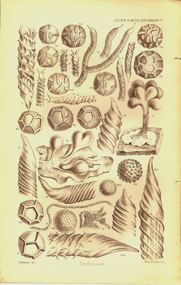
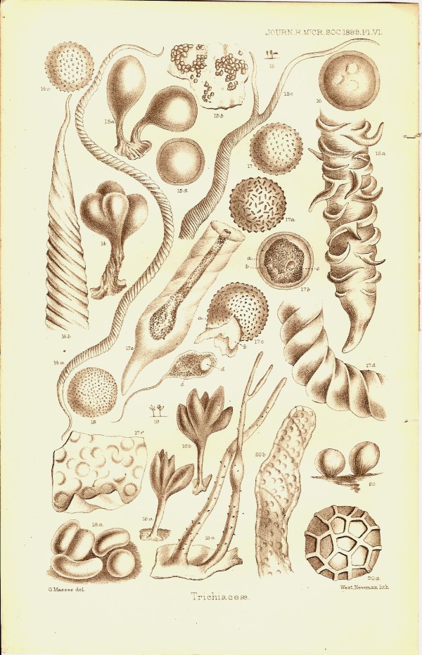
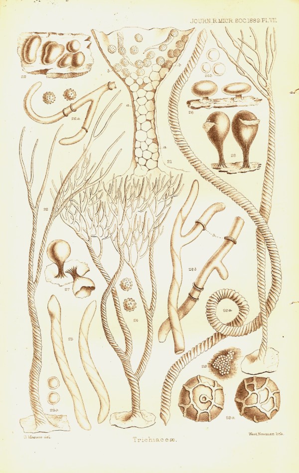
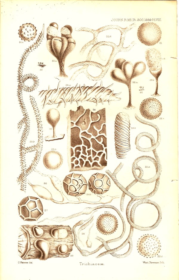
Two or three years ago, I bought a number of early issues of the Journal of the Royal Microscopical Society at a very reasonable price. This afternoon I pulled one off the shelf at random and started browsing in it–always a delightfully dangerous diversion, since inevitably I find something I want to write about. This particular copy is from June 1889. I have always delighted in Victorian titles of books and journals. Journal of the Royal Microscopical Society wasn’t enough; no, then we get a semicolon and the subtitle “containing its transactions and proceedings and a summary of current researches relating to ZOOLOGY AND BOTANY (principally Invertebrata and Cryptogamia), MICROSCOPY, &c.” Imagine the poor librarians who had to try to fit such a title in a 3 inch blank on an index card. This is a handsome and substantial publication consisting of 151 pages not including the cover pages and advertisement pages; in fact, it is a most impressive journal that covers a broad array of reports of current researches and I’ll give a summary in a moment. However, the opening piece is an article which runs from p. 325 to p. 359 and this is Part VI of “A Revision of the Trichiaceae” by George Massee. (See, I’m not the only serial scribbler.) These organisms are Mycetozoa or Myxomycetes and the ones under discussion are illustrated by 4 marvelous plates of drawings which are so intriguing that I’ll include them here for you to enjoy. I should tell you, in case you haven’t come across these utterly fascinating and wildly bizarre organisms, that they are popularly known as slime molds. These are the creatures of science fiction. Their life cycles are so strange that both botanists and zoologists used to claim them. Slime molds aren’t really molds, although in one stage of their development, they produce spores from fruiting bodies that do somewhat resemble miniature fungi but, in addition, they can have an amoeboid state which is multinucleate and which can achieve a size of over 2 feet. When one produces fruiting bodies, they release spores and the cycle begins all over again. The designation “slime mold” prompts one to think of something repellent, but in fact, many species have developmental stages that are quite lovely and some are quite colorful as well, but since the plates are black and white, that does not show up here. So here, without further ado, are the plates.
Several years ago, my wife had a number of African violets that needed trimming up and I, ever on the lookout for new material for my lab, volunteered to trim them. I was curious about what sorts of results I might get if I put some of the leaves in medium-sized culture dishes, covered them with water, and allowed them to ferment–perhaps some exotic protozoans might appear. I prepared four dishes, set them on a shelf and rather forgot about them for several weeks. Upon remembering them, I was afraid that they would have transformed themselves into slimy, green, gelatinous, smelly messes, but not so. Violet leaves are apparently not subject to rapid decay and when I opened the dishes, I was I admit rather disappointed–quite a lot of white, fluffy mold and, in the end it turned out to be quite attractive under the microscope. However, I was looking for protozoa, so my examination was casual, too casual; I want to try this experiment again and try to get some images. The first time I did this I didn’t have a microscope camera. When I went back to the dishes for a second look, I noticed some odd little greenish-grey globular structures on some of the leaves–slime molds! I was enormously excited, since I had never seen one in the flesh, as it were. I have a few commercial slides of some of the fruiting bodies which are fascinating, but these strange little growths were real micro-treasures to me. I took notes; but no camera, so no images. So, I have resolved to trim violets again and if I get any results before this essay appears, I’ll add some images. Slime molds are actually fairly common, but tend to be overlooked because most of them are quite small and they live in habitats we often neglect, such as the undersides of logs in damp forests. I urge any amateur who is interested in micro-life to spend some time trying to find and observe slime molds. Many of you know that wonderful series of “How To Know The ______” books, such as Jahn’s How to Know the Protozoaor Prescott’s How to Know the Freshwater Algae. Well believe it or not, there is in that series a How To Know The Slime Molds and a very nice reference it is.
After this major article, there is an extensive series of brief reports which constitute a “Summary of Current Researches”. The first major heading is Zoology with two primary subheadings: Vertebrata and Invertebrata. Vertebrata has three secondary subheadings: Embryology, Histology, and General. Just to give you an idea of the character of these summaries, which run from a quarter of a page to two pages, I’ll give you a few titles. Under Embryology, there is E. Ray Lankester’s “Structure of Amphioxus laceolatus”. Lankester did an important multi-volume work of zoology. He was a distinguished zoologist who was knighted in 1907. For those of you, not familiar with Amphioxus, it is one of those wonderfully intriguing transitional organisms. It looks rather like a tiny fish without fins and it does indeed have primitive notochord or “backbone”.

There are a number of groups of organisms that evolved on that fascinating edge between the invertebrates and the vertebrates. Amphioxus is described as a cephalochordate in part because it has a distinct head region. Then there are Urochordates and Protochordates. The fossil record shows that on a number of occasions, life attempted to cross this bridge from squishy, spineless creatures (such as politicians) to those with a backbone. Some of these experiments failed and those organisms have been long extinct. Some of them survived, but only got half way. The sessile tunicates, for example, evolved in such a way that in the larval stages, they possess a notochord. However, when the larvae attach and begin to transform to the adult stage, the notochord disappears. Ane even when we clearly get to vertebrate territory with the fishes, we discover two distinct strategies: the cartilaginous fishes, such as sharks, and the bony fish which are far more numerous.
In the Histology
section, there is a piece on the “Structure of the Cell and Phenomena of
its Division” and in the General section a page on “Fresh-water Fauna of
Greenland.” The researches being reported here are real science and not
the popular science or amateur natural history of the sort that we found
so abundantly in the previous series: A Trip Into The Past.
The reports on
research regarding invertebrates is even more impressive and covers
cephalopods, gastropods, lamellibranchs, tunicates, bryozoa, insects,
arachnids, crustacea, annelids, nemathelminths, platyhelminths,
echinoderms, coelenterates, porifera, and
protozoa.
The botany section
is also highly diverse and includes reports of work on cell structures,
tissues, organs, on physiology, on algae, fungi, slime molds, diatoms,
desmids, and bacteria.
The final section is
on microscopy and is divided into two major subsections: 1) Instruments,
Accessories, &c. and 2) Technique. I’ll mention just a few of the
titles from the Invertebrate and Microscopy sections which caught my
eye.
1) “Hermaphroditism
of Aplysiae.” These biologists all seem preoccupied with sex, which
reminds me of the old joke about the man who goes to a psychiatrist and
the psychiatrist decides to give him the classic Rorschach Ink Blot test.
He tells the patient: “Now, when you look at each image, I want you to
tell me the first thing that it makes you think of.” The doctor holds up
the first ink blot image and the patient says: “That’s a man and a woman
having sex.” He holds up the second image and the patient says: “That’s
two couples having sex.” He holds up a third image and the patient says:
“Oh, that’s an orgy.” The psychiatrist looks over at the patient and says:
My, you certainly are obsessed with sex.” And the patient replies: “Me?
You’re the one with all the dirty pictures!”
As a consequence of
our perspectives regarding human sexuality, we often forget that this also
is an area in which nature constantly experiments. Sexuality is
essentially a series of processes to preserve the strongest genes and to
select ones that can adapt to new conditions. Whatever the biochemical and
biophysical dynamics of life are, one thing is clear; it is absolutely
ruthless in its struggle to survive.
It is astonishing for us to think that even protozoa exhibit sexual or “mating” types and that in some termite symbiotic flagellates there are as many as five different sexes! There is a fairly wide range of invertebrates which are hermaphroditic, that is, they are both male and female. This is not uncommon among the mollusks and Aplysia or the “sea hare” is a highly evolved mollusk with only the remnants of a shell. The sea hare is of special interest to biologists because of its giant ganglia. It derives its common name from the fact that it has two projections on its head which do give it a vague resemblance to a rabbit.
In the section on Protozoa, there is a very brief note titled: “Holotrichous Infusoria parasitic in White Ant” with a reference to a 3 page article by W.J. Simmons in The Microscope, ix; (1889) pp. 53-55. This item is of interest because of the terms in the title. Generally the term “holotrichous” which means “having cilia over the entire surface”is applied to ciliate protozoa. “White ants” often used to be a description for termites. “Infusoria” in the 18thand 19thCenturies was often a catchall term that included not only protozoa, but a fairly wide variety of microscopic aquatic life forms. The note states that the author seems in doubt about the morphology of the organism because it is constantly changing shape. I don’t know if the original article had any drawings that might help identify the organism and, if I weren’t so lazy, I might check. Instead I’ll indulge myself in a little speculative enterprise. Let’s assume that the white ants were indeed termites. It was reported that the organisms were in the lower intestines and were extremely abundant. Now, since the term “infusoria” was used rather than “ciliate”, it’s possible that these organisms were hypermastigote flagellates and might even have been of the genus Trichonympha which are very abundant in the gut of the termite and possess a highly flexible membrane so that they can indeed present major problems in trying to determine their morphology. These are sizeable flagellates which possess hundreds, if not thousands, of flagella. If they were indeed Trichonympha, then they are not parasites, but symbionts which, in fact, are necessary for the survival of the termites, since they cannot metabolize cellulose and depend upon the protozoa to produce byproducts which the termites can use for nutrition. I mention these factors to illustrate how shifts in the meaning of terminology can significantly alter the significance of a particular account. I decided to overcome my lethargy and will make an effort to have the university library locate a copy of the article if possible and then let you know if my little thought experiment holds water.
RESULTS :
Ah, the miracles of
modern technology. Five days ago, I contacted a reference librarian at the
university by telephone and he reported that the library had some earlier
issues of this journal, but not the 1889 one. I then went online to the
InterLibrary Loan department and filled out a request form which I
e-mailed to them. They did a search of other libraries and then today–only
5 days later–they electronically sent me the pages which I had requested
and I printed them off and have them sitting here before me–all without
leaving the house. What a time to be a scholar!
So, what did I find
out?
1) Mr. Simmons was
prompted by the work of Professor Leidy, a distinguished biologist and
director of the biology program when founded at the University of
Pennsylvania, who did work on the American white ant which, it becomes
clear in the course of the article, is indeed a termite. Mr. Simmons,
however, was working on white ants from Calcutta, so we don’t know whether
or not he was examining the same species.
2) He comments: “The
alimentary canal of these ants...teem in its lower portion with parasites.
Although moniliform and ciliate and non-ciliated organisms, bacteria,
spirilla and nematoid worms were found, the present paper is devoted to
the Holotricha.”
This list of organisms is consistent with what can be found in the gut of termites.” “Holotrichous” means covered all over with cilia, except in this case the organelles described are really flagella and indeed it is very likely that this is a symbiont and not a parasite. Apparently I was mistaken in one part of my conjecture for the article states: “No contractile vesicle has yet been detected, a feature this parasite shares in common with the genera Trichonympha and Pyrsonema of Leidy.” So, Simmons is treating his organism as being closely related to these two genera of flagellates. In an important way,, this instance poses a classic dilemma confronted by 19th Century microscopists doing taxonomy and so I would like to take a closer look at some of the details.
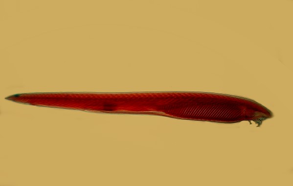
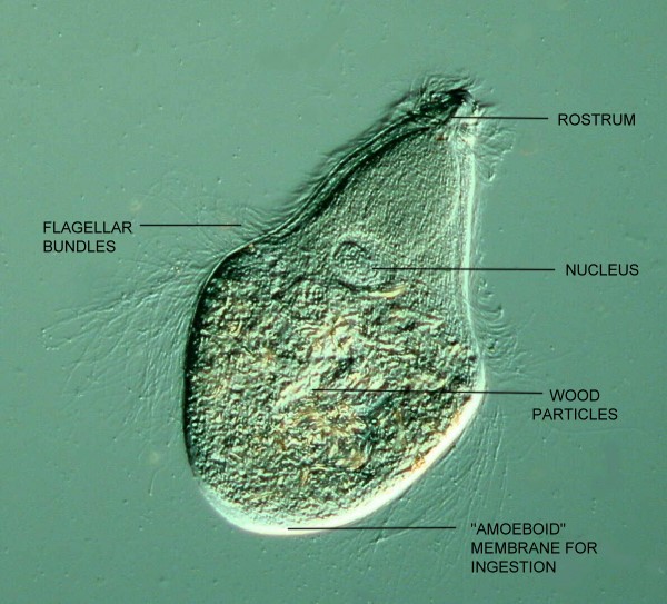
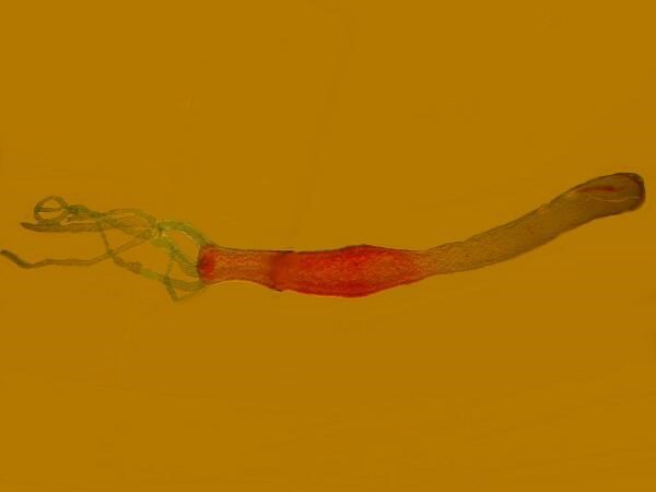
3) Unfortunately no drawings of the organism in question are provided and verbal descriptions are often of only limited use as law courts have demonstrated repeatedly from testimony of witnesses who describe an alleged perpetrator of a crime as somewhere between 5 foot and 6 ½ foot tall, with blue, black green or gray eyes, clean-shaven with a moustache and full beard, and blond, gray or red hair. In spite of the fact that Simmons says that this organism is not Trichonympha, I am still inclined to suspect that the organism in question might still be Trichonympha or a very close relative–however, I admit I might well be wrong. But let me show you a couple of pictures of Trichonympha campanula and then we’ll consider Simmons’ account further.
The description of Simmons’ organism continues:
“The body frequently shows parallel spiral markings which may indicate the position of the cilia, or a ridge surface...There is a distinct and large nucleus, of circular form, the general location of which is central, though it may be nearer one or other end of the body...The body is generally gorged with food, identical in appearance with the contents of the alimentary canal of the termites in which the parasites occur...The ‘mouth-parts’ of the organism is a hyaline cap surmounting a narrow tube, probably pharyngeal, which is in most cases located at the anterior extremity. It does not occur in all the parasites; and moreover, in some instances the cap is replaced by a minute hyaline sphere.”
Let’s pause for a moment to examine these remarks. The only aspect that is not commensurate with thinking that this being a Trichonympha-like organism is the remark about the “mouth parts”.
As it turns out, this issue of the “mouth parts” is especially interesting. Let me quote one more brief passage before discussing its importance.
“Simmons expresses himself provisionally as to these organs being mouth-parts, never having seen food particles pass into the mouth; nor through the pharyngeal tube, nor detected them in its immediate neighborhood; indeed, the dimensions of some of the particles have been such as to preclude the possibility of their having passed down the tube, unless it be dilatable. From the identity of the food particles in the parasite with those in the intestinal organs of the termite, we must infer with Leidy that an oral aperture exists.”
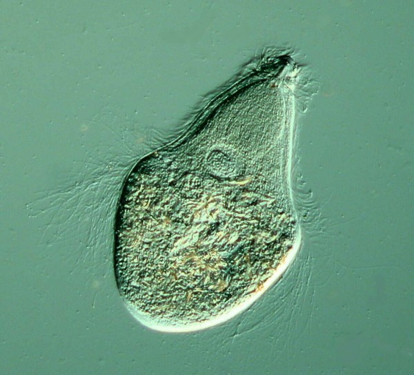
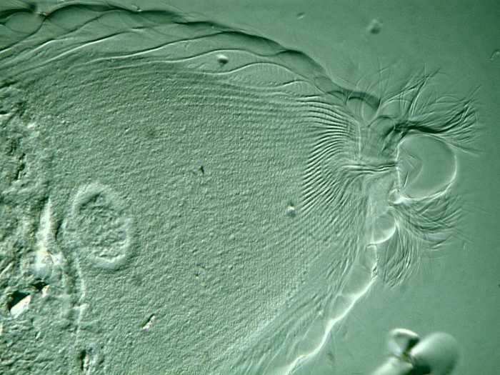
Such an inference is dubious and Trichonympha is a perfect example to show why. The general shape of the organism with its distinctive rostrum strongly suggests that feeding should take place there at the anterior end. SURPRISE! Feeding takes place at the posterior end where there is an area with a clear membrane around the body as is visible in the 2nd image below.
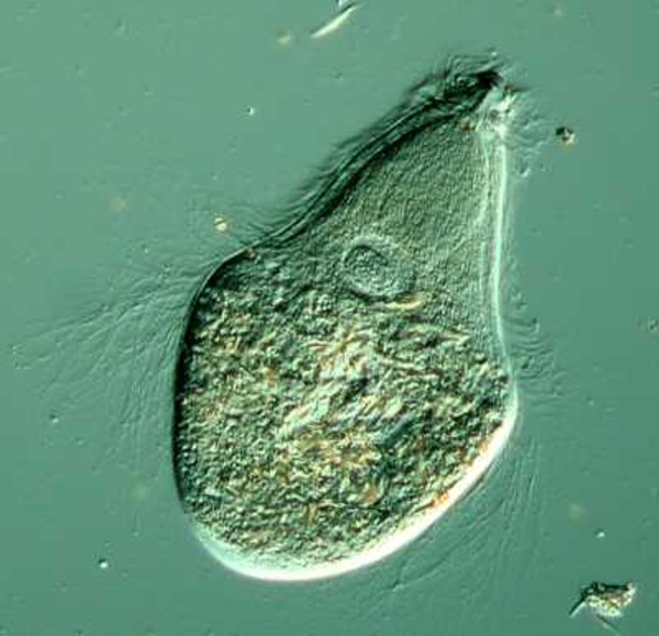
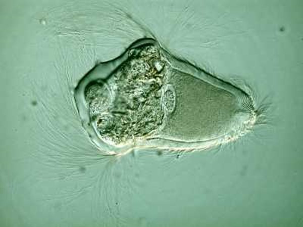
In the second image, you can see the irregularity of the area and, as it turns out, Trichonympha feeds in an amoeboid-like manner projecting pseudopodia-like extrusions from this area of the membrane. Mother Nature is such a trickster and I think that’s why virtually every mythological system I know of has a trickster god, goddess, or demi-god.
In one sense, we shouldn’t be too surprised, since there are at least two groups of amoebae with flagella and there are flagellates with amoeboid stages.
Simmons seems convinced that this organism is something other than Trichonympha and he may well be right. There are some extraordinarily weird organisms living in termites. I would add, however, that the contortions of which Trichonympha can undergo, make it very difficult to identify it from certain perspectives. All of this underscores the importance of having detailed drawings in addition to photomicrographs and/or video.
In the section on Fungi, there is a report on the researches of G. Dupetit “Toxic Principles of Fungi”. Judging from this issue of the journal, there was widespread interest in fungi in the late 19th Century. We know that at least since the beginning of recorded history, fungi have been gathered as food and as hallucinogens and sometimes have even snuck into the food supply unbeknownst to the consumers with devastating consequences. Anyone still living, who has collected “edible” mushrooms knows that considerable caution and some modest expertise are essential for survival. There are extraordinarily complex chemicals in some fungi that can interact with the human body in bizarre and sometimes unpredictable ways. For example, some mushrooms that are ordinarily quite benign can turn very nasty if the person eating them is also imbibing alcohol. There is also the risk to the neophyte collector that some very dangerous mushrooms are visually very attractive, Amanita muscaria, for example. In the mountains to the west of us, I have found beautiful specimens with the cream colored stalk, the bright red-orange cap with its white stipples scattered around the surface. It is lovely, but lethal. It is commonly known as the fly agaric. Other species of Amanita are also quite dangerous, such as, Amanita verna, the Destroying Angel, and Amanita virosa, the Death Cap, as the ingestion of just a half of one cap can be fatal to a vigorous adult. Some foolhardy sorts have tried using minute quantities of these fungi to induce hallucinations. Many fungi contain very powerful alkaloid chemicals which can produce bizarre symptoms.
I mentioned that sometimes fungi get into the food supply without our notice. A classic example of this is ergot which grows on rye. It too contains a powerful alkaloid and when ingested causes contraction of blood vessels, including those in the brain. Among its symptoms are nervous spasms, convulsions, wild uncontrolled movement, and gangrene. In Medieval times it was known as ignis sacer, “holy fire” and also came to be known as Saint Anthony’s Fire. This phenomenon became associated with both the plague and with witchcraft. Ironically in the 20th Century, there have been outbreaks of what at first appeared to be ergot poisoning, but were later discovered to be due to toxic mercury compounds which were used to treat grain to keep it from being infected with fungal growths. Twentieth Century medical science extracted this compound and found ways to make it useful, especially in the treatment of migraine headaches.
In the section on Microscopy, Instruments, Accessories, &c., there are a number of especially intriguing items, including a letter from Darwin written to Sir Richard Owen in1848, regarding his microscope, this being of sufficient interest that I will quote it in its entirety.
My Dear Owen,
I do not know whether your MS instructions are sent in; but even if they are not sent in, I dare say what I am going to write will be absolutely superfluous, but I have derived such infinitely great advantage from my new simple Microscope, in comparison with the one which I used on board the ‘Beagle,’ and which was recommended to me by R. Brown, that I cannot forgo the mere chance of great advantage of urging this on you. The leading point of difference consists simply in having the stage for saucers very large and fixed–mine will hold a saucer three inches in inside diameter. I have never seen such a Microscope as mine, though Chevalier’s (from whose plan many points of mine are taken), of Paris, approaches it pretty closely. I fully appreciate the utter absurdity of my giving you advice about means of dissecting; but I have appreciated myself the enormous disadvantage of having worked with a bad instrument, though thought a few years since the best. Please to observe that, without you call special attention to this point, those ignorant of natural history will be sure to get one of the fiddling instruments sold in shops. If you thought fit, I would point out the differences which, from my experience, make a useful Microscope for the kind of dissection, of the invertebrates, which a person would be likely to attempt on board a vessel. But pray, again believe me that I feel the absurdity of this letter, and I write merely from the chance of yourself possessing great skill and having worked with good instruments, may not possibly be fully aware what an astonishing difference the kind of Microscope makes for those who have not been trained in skill for dissection under water...
Ever, my dear Owen,
Yours sincerely,
C. Darwin.
P.S.–If I do not hear I shall understand that my letter is superfluous. Smith and Beck were so pleased with the simple Microscope they made for me, that they have made another as a model. If you are consulted by any young naturalists, do recommend them to look at this; I really feel quite a personal gratitude to this form of Microscope and quite a hatred to my old one.
Professor Owen was a member of the Royal College of Surgeons which, in part, explains Darwin’s reluctance in recommending a dissecting microscope to him.
There are two points in particular that I find of special interest in this letter. I find it mildly amusing that Darwin rather blithely assumes that there is going to be a significant demand for microscopes to perform dissections of invertebrates on shipboard. It is true that during this period the wide-ranging interest in natural history resulted in numerous ships having a naturalist aboard and, of course, there were also major ventures to explore and collect sea life, such as, the Challenger Expedition. However, the majority of the ships were cargo ships and the owners were primarily interested in sizeable exotic specimens that were in high demand in Great Britain, Europe, and America. Doing minute dissection under a microscope was a rarity outside of major scientific expeditions.
Clearly Darwin himself was highly adept at doing micro-dissection; I’m not sure that I would want to try that on a ship that could start pitching and rolling at any time. Another indication of this expertise and his keen powers of observation is the treatise on barnacles which he produced. He spent 8 years dissecting and describing barnacles in a work which is still a classic.
Most serious students of biology today have a general knowledge of the major aspects of Darwinian theory, but few of them have an idea of the remarkable range of his interests and writings. Almost everyone knows of The Origin of Species and The Voyage of the Beagle, although some make pronouncements about them without having read them. They have read summaries, encyclopedia articles, or someone’s brief book presenting the outlines of Darwinian views. To me, this is like buying a CD of 100 of the World’s Greatest Classical Masterpieces–the producers select out the great melodies for you (and eliminate all the “dull” parts). It’s basically the same kind of objection I’ve always had to speed reading which Woody Allen expresses so well: “I took a speed-reading course and read War and Peace in twenty minutes. It involves Russia.” Many contemporary students have no conception that Darwin’s interest in natural history produced such works as:
1) On Structure and
Distribution of Coral Reefs
2) Power of Movement
in Plants
3) Animals and
Plants under Domestication
4) The Descent of
Man and Selection in Relation to Sex
5) The Formation of
Vegetable Mould Through the Action of Worms with Observations on their
Habits
6) Different Forms
of Flowers on Plants of the Same Species
7) Insectivorous
Plants
8) The Expression of
the Emotions in Man and Animals
9) Climbing
Plants
10) The Various
Contrivances by which British and Foreign Orchids are Fertilized by
Insects
I was also interested to note that in the letter Darwin makes reference to those “fiddling instruments sold in shops” which suggests the sorts of “microscopes for children” sold these days.
The other item in Darwin’s letter that I find both interesting and amusing is the postscript. Darwin is deferential to Owen, almost to the point of obsequiousness, but then becomes very direct and assertive that Owen should recommend this new microscope to young naturalists. I also find quite delightful his description of his relation to his microscopes–his “personal gratitude” to the new one and that he had developed “quite a hatred to my old one.” A feeling that I suspect quite a number of us have had.
At the back of this issue, there are 4 ½ pages of advertisements (the other ½ page being a listing of the members of the Council of the Royal Microscopical Society). Almost all of them have to do either with microscopes or cameras or slides or, in one case, a book. There are, however, two notable exceptions. The first is an advertisement for “The Luxury of Pure Water” from W. & J. Burrow, The Springs, Malvern “Patentees of ‘the Slider’ Wine Bins. In fact, they recommend their sparkling water as being “corrective, healthful, and delicious either alone or with Wine or Spirit”. In addition to sparkling water they sold still water, seltzer, soda, Potash, Lithia, “and other waters of exceptional purity.” It’s nice to know that the current mania for “natural” and “pure” bottled spring waters is well over a century old. Here, I am afraid, I’m in agreement with W.C. Fields, who asked why anyone should spoil good Spirits by adding water and with Mark Twain who said: “Water, taken in moderation, cannot hurt anybody.”
The other advertisement is for:
EPPS (GRATEFUL COMFORTING) COCOA
The entire advertisement consists of an endorsement from, of all places, the Civil Service Gazette (British, of course). Here it is in all it’s 19th Century glory for your delectation:
“By a thorough knowledge of the natural laws which govern the operations of digestion and nutrition, and by a careful application of the five properties of well-selected Cocoa, Mr. EPPS has provided our breakfast-tables with a delicately-flavoured beverage which may save us many heavy doctors’ bills. It is by the judicious use of such articles of diet that a constitution may be gradually built up until strong enough to resist every tendency to disease. Hundreds of subtle maladies are floating around us ready to attack wherever there is a weak point. We may escape many a fatal shaft by keeping ourselves well fortified with pure blood and a properly nourished frame.”
Now here’s a health plan that George Bush and Sir Humphrey Appleby could agree on!
Anyone who is interested in antique slides knows of the firm W. Watson & Sons which was established in 1837. Now, in this journal 52 years later, they advertise a “Classified List of Stock of 40,000 first-class Objects, illustrating every branch of study.” The Watson slides have a fine reputation and the few I have seen are of high quality. It is tempting to fantasize having written them a letter saying, “I’ll take one of each.”
The range of subject in 19th Century slides is simply amazing and quite wonderful. Consider these micro-marvels offered in this advertisement.
1) “Section through the Bud of Lily, showing Ovary,, Anthers, Pollen-grains, and Petals in situ” for a mere 2 shillings. This was before the British converted the shilling to cents, thus ruining their economy and their wonderfully bewildering currency. If you’ve seen slides of sections of lily, you know what elegant objects they are and to have 4 different aspects present on a single slide is especially wonderful.

2) “Horizontal Section through Foot of Human Embryo, showing commencement of ossification in Metacarpal Bones, very fine, 3s. 6d.” Today the use of human embryonic tissue for any purpose has become highly controversial. I just looked at a catalog from a major scientific supply house and was unable to find any slides of human embryonic material. How major a supply house?–well, their catalog is over 1,300 pages.
3) “Section through entire Human Eye, 5s.” We tend to take our eyes for granted and yet they are an extraordinary accomplishment in the process of evolution. About every year, I have to get a new prescription for my glasses or else grow longer arms. Light-sensitive organelles developed as early as the protozoa.
At much later evolutionary stages, we find eyes of extraordinary sophistication and acuity in cephalopods, birds, and humans.
4)”Anatomy of Leaf, nine pieces on one slide, 2s. 6d.”
5) Typical examples of Blood from Man, Bird, Fish, Snake, and Frog, on one slide, 5s.”
These two types of slides are especially valuable for the instruction of students; the leaf slide gives a sense for how the various parts fit into the composite and the blood slide is excellent for comparative studies.
6) “Type slide of fifty British Foraminifera, name photographed beneath each specimen. £1 1s.”
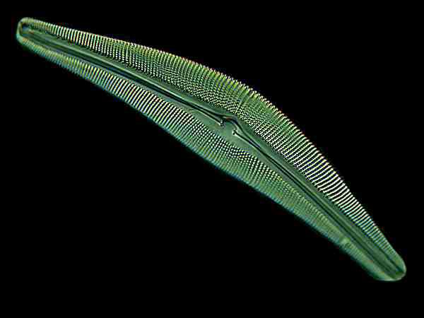
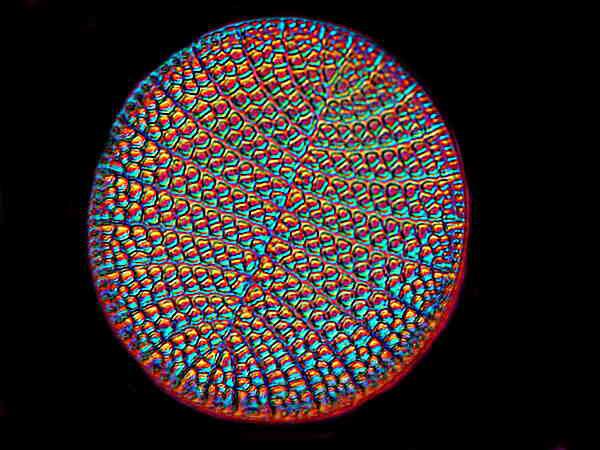
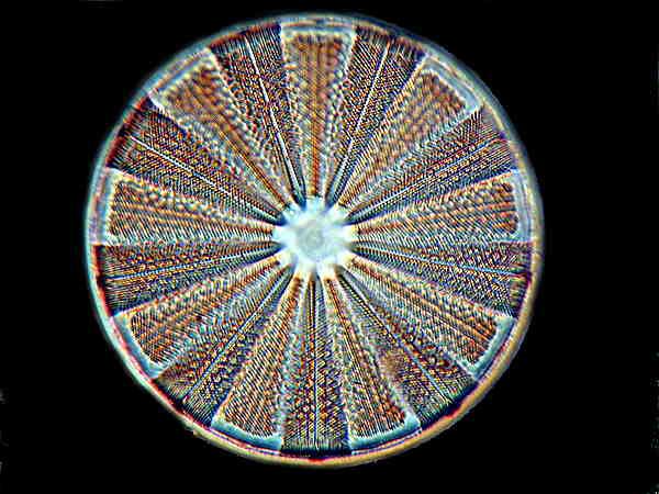
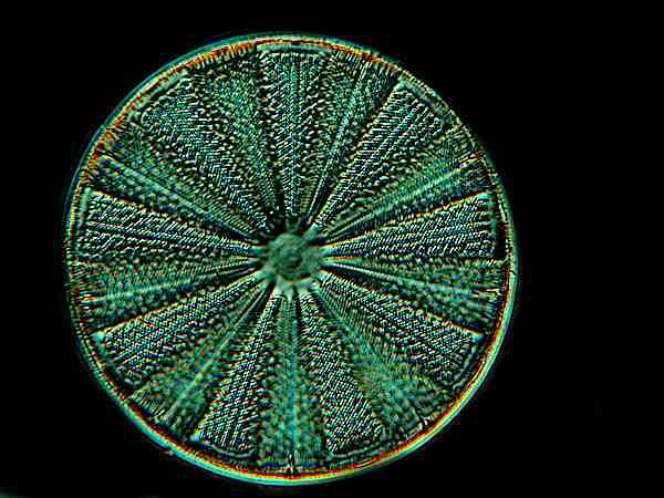
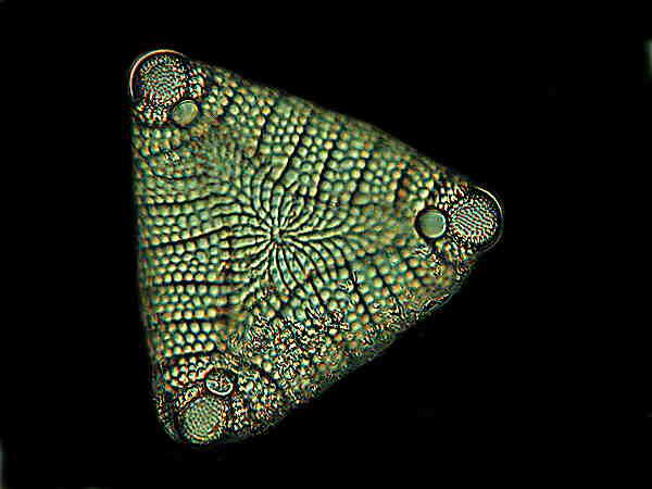
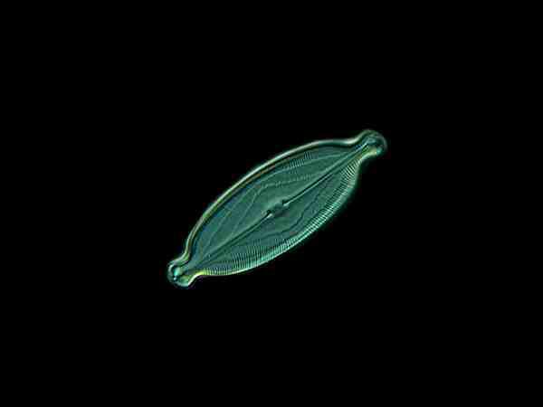
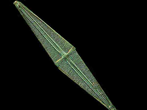
7) “Type slide of about seventy Diatoms from Oamaru, N.Z. 10s 6d.”
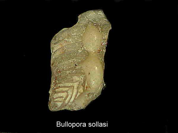
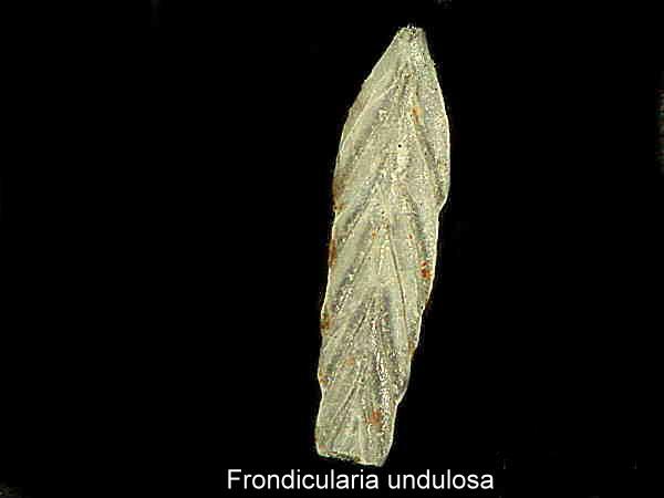
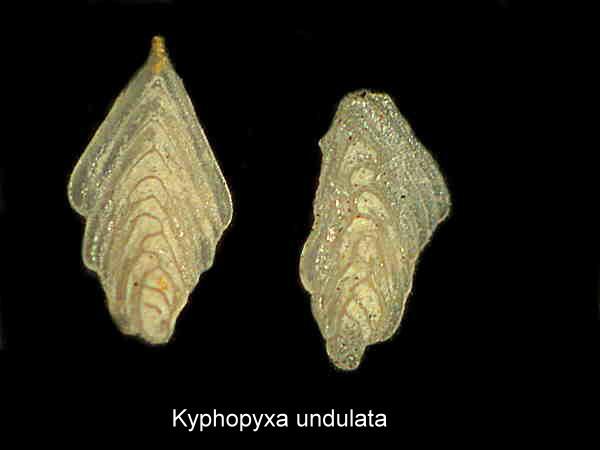
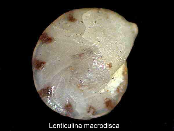
Type slides are highly sought after and are a very valuable resource, especially those which included the identification on the slide itself. Most type slides come with a separate list identifying the objects, but such lists are all too easy to lose. For those of you who may be unfamiliar with the foraminifera (or forams, for short), they are marine amoebae which form elegant, and sometimes bizarre, microscopic shells which are usually calcareous. There are literally thousands of species and they have been extensively studied, not only for their intrinsic scientific and aesthetic character, but for their economic significance as well. Prior to the rather recent development of new technologies, core samples containing forams were widely used as indicators for the location of oil deposits. I’ll give you a few examples of foram shells here.
The type slide of diatoms from Oamaru, New Zealand is also of special interest since this site has produced and still produces some especially magnificent specimens. Again, in case you haven’t come across diatoms, these are the microscopic shells of organisms which used to be classified as plants, but are now assigned to the Kingdom Protista (or Protoctista). These shells are composed largely of silica (glass) and they are capable of a slow and puzzling motion. The current theory for accounting for this motion is streaming protoplasm around the outside of the shell. These shells are among the most beautiful objects to be found in nature. Here are a few examples.
8) “Hydra vulgaris, 1s. 6d.” Hydra, one of the very few freshwater coelenterates, is fortunately common (vulgaris) and a wonderful creature to study. It has fascinated aquatic biologists back as for as Trembley’s study published in 1744. One can learn an enormous amount of biology from this one organism. Although it seems quite primitive, a simple tube-like stalk with tentacles, but these tentacles have four different kinds of nematocysts or “stinging cells” which capture food, such as, the water flea Daphnia and they push it into the mouth located at the base of the tentacles. Furthermore, though it consists of only a relatively small number of cell types, it can produce ovaries, spermaries, or reproduce by budding, so that projecting out from the stalk you may see one, two or even more developing hydras. Moreover, these amazing little creatures are capable of regeneration.
9) “Very beautiful slide of Eggs of Butterflies and Moths, &c., arranged in a group, 10s. 6d.” I used to stalk butterflies, following them around, trying to catch them in the act of laying eggs so that I could collect them and mount them. They are indeed lovely objects. However, I was never successful and it is not a boost to one’s ego to be repeatedly outsmarted by insects, so I finally gave up. Last summer, I was sitting out on our tiny side porch, when I noticed a tiny cluster of small objects–Eureka! Well, sort of. They were the vestiges of egg cases. The critters had hatched and flown. Nonetheless, these were still interesting objects. Imagine a Roman stone urn made of a translucent parchment-like substance and also possessing a tiny hinged lid of the same material. Quite wonderful, but even so, I would still like to have a slide of arranged butterfly and moth eggs such as the one that W. Watson & Sons sold. In fact, I wish I could buy all nine of these slides that I have described for the prices they listed for them.
All comments to the author Richard Howey are welcomed.
Editor's note: Visit Richard Howey's new website at http://rhowey.googlepages.com/home where he plans to share aspects of his wide interests.
Microscopy UK Front
Page
Micscape
Magazine
Article
Library
Please report any Web problems or offer general comments to the Micscape Editor .
Micscape is the on-line monthly magazine of the Microscopy UK website at Microscopy-UK .