PLANARIA: THE DAY WE INVITED THEM FOR LUNCH
Manuel del Cerro, Pittsford, NY
Marilu DeCoste, Princeton, NJ
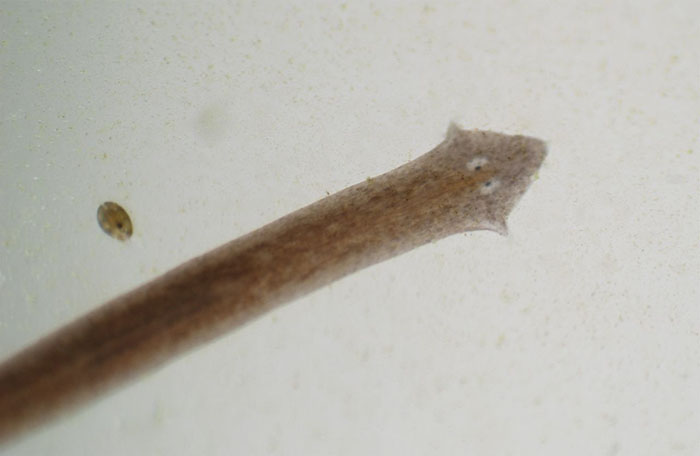
Figure 1. A Planaria cruising on the micro-aquarium wall; an ostracod is seen close by. The field in this and all pictures, except figure 7, is 1.1 cm across.
INTRODUCTION
Planaria (a name we use both as singular and plural) is a worm frequently found in ponds and small streams. It is a flat worm (phylum Platyhelminths), that has been the object of numerous studies for some two hundred years. Biology students and molecular biology researchers, amateur and professional microscopists, all have been interested in Planaria. Today, Planaria continues to be an object of great interest in stem cells research and into the search for means to limit tumor growth in humans and animals.
The extraordinary regeneration powers of Planaria has attracted much attention. The worm can be cut transversally or longitudinally and the fragments regenerate full individuals. Variations of the procedure allow the production of worms with multiple heads. It was found that Planaria exhibit definite memory characteristics. They can also develop conditioned reflexes (Block and McConnell, 1967) and these reflexes can be retained by the two individuals created be surgically dividing the worm. The claim was even made that a conditioned reflex could be perpetuated by grinding conditioned Planaria and feeding the mush to other Planaria. This claim sent investigators around the world into a frantic search for the “memory molecule.” Unfortunately, the result was never independently reproduced (Rilling, 1996).
MATERIAL AND METHODS
Subjects and Habitat. Our “Micro-aquarium” has been described previously in this publication (del Cerro, 2007). A year ago, a water sample obtained at Powder Mills Park, a county park located off route 96, in Pittsford, NY, southeast of Rochester, NY, was introduced into the micro-aquarium. Days later a few Planaria were seen exploring the glass walls of the container; the population has expanded considerably since. It is worth noting that Planaria are hermaphroditic but not self-impregnating; they reproduce by both sexual pairing and non-sexual means (Buchsbaum, 1938).
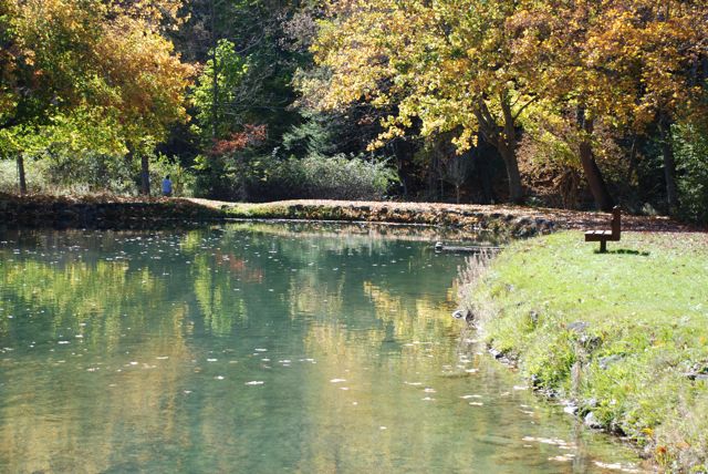
Figure 2. An artificial pond in Powder Mill Park. The water sample containing our original Planaria stock was collected from the inlet to this pond.
Photographic technique. An AF Nikkor zoom lens, 35-80 mm, was reverse-mounted on the body of a digital D80 Nikon camera using a Nikon BR-2 ring as an adapter (Cooper and Abbott, 1979). This set up provides a range of magnifications that goes from a field as wide as 4.5 cm with the lens zoomed to 80 mm focal length, to as narrow as 1.1 cm when set to 35 mm of focal length; the latter was the preferred option for the pictures illustrating this note. The male bayonet thread of the ring fits perfectly into the female bayonet thread of the camera body but the functional compatibility is limited so that the camera will only operate in the manual mode. This is not a serious limitation for close up work given that exposure time, f opening, focusing and zooming can all be manually controlled.
What’s for dinner? Planaria seem to enjoy any form of tropical fish food. In this case we opted for placing close to the front glass panel of the micro-aquarium grains of small-size floating pellets for Central American tropical fish.
The tools of gastronomic pleasure. At difference with the overwhelming majority of animals species, Planaria does not have the mouth located in its head. The orifice called the “mouth” is instead located on the lower surface of the body, about half way between the tip of the head and the tip of the tail. A further oddity is the fact that food is not ingested through the mouth. A tubular structure called the “pharynx” normally kept inside the body is projected out through the mouth at feeding time. The pharynx works like a straw, and the Planaria ingests its food as it were sucking a “slurpee” (Wikipedia, 2009). What happens after? The pharynx brings the food into a “gastrovascular cavity” to which it is connected. The gastrovascular cavity is shaped like a tuning fork, with the stem directed towards the head and the arms towards the back, running parallel to the pharynx. It has multiple out-pockets that become visible in the recently fed animal; several of these structures are visible in the figures below. Once in the gastrovascular cavity the food particles are taken in by ameboid cells lining the cavity; digestion of the food particles is intracellular. We will go no further in describing the anatomy of Planaria but strongly recommend searching for more information on this fascinating animal either in chapter 10 of Buchsbaum’s book (1938), or in the Planaria entry in Wikipedia (2009); both make pleasant and informative reading.
OBSERVATIONS
Lunch time. The food pellets were introduced on 10 May 2009, at 1210 hours local time. The first Planaria arrived to the proximity of the pellets by 1220 hours (figure 3) and reached the pellets almost immediately (figure 4); it explored the pellet for a moment and then projected its pharynx out from its body and onto the pellet (figure 5).
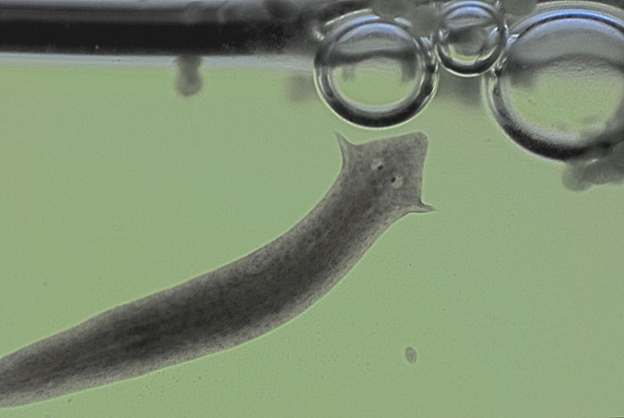
Figure 3. A Planaria that was nearby came searching for the pellets (not visible in this field) that were floating on the water surface, to the right of the air bubbles. The field is 1.1 cm wide.
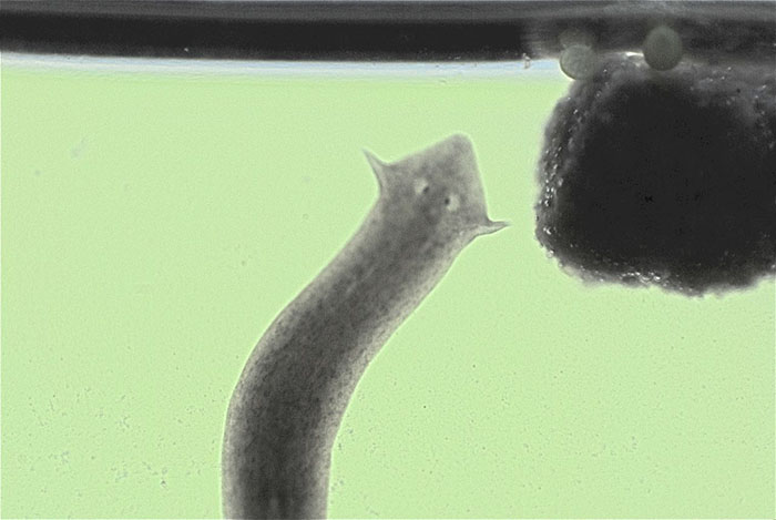
Figure 4. The same Planaria is about to contact one of the food pellets. The body is heavily pigmented, so that little of the internal structure can be discerned except the eyes. The horizontal line separating the light from the dark areas of the picture is the water surface. The field is 1.1 cm wide.
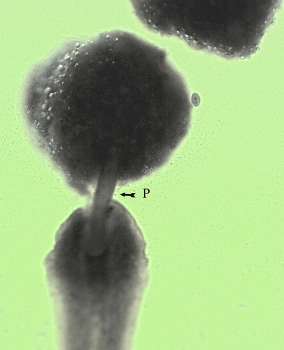
Figure 5. The fully extended pharynx appears as a tube linking the body of the Planaria to the food pellet that is now fully immersed. Small air bubbles cover the surface of the pellet. An ostracod swims to the right of the food pellet.
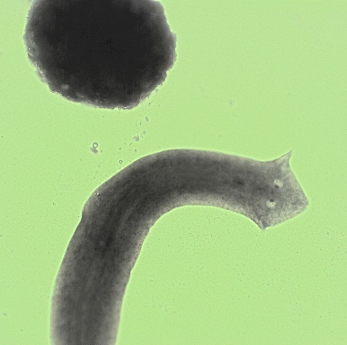
Figure 6. Now well fed, the Planaria abandons the food pellet.
By 1230 hours the Planaria was feeding itself and continued doing so for about 20 minutes. In the meantime, many Planaria were emerging from the mat of algae and debris covering the bottom of the aquarium and were gliding fast on the walls on all sides of the aquarium. Their navigation towards the pellets was quite precise, there was little meandering and we have seen no individual equivocating its way. They must be detecting and following with high proficiency a signal gradient, be it gustatory or olfactory. There was a single pellet floating near a cluster of four other; Planaria paid little attention to the single pellet, favoring exclusively the cluster. Again, this suggests that they detected and followed the strongest signal. By 1300 hours, there was frantic activity around the pellets with new individuals arriving as the first ones were leaving (figures 7 and 8).
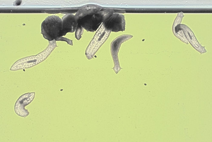
Figure 7. As many as ten Planaria are feasting on the food pellets. Two are on top of the pellets and are difficult to see except for the bump that their bodies produce on the water surface. Notice the difference in pigmentation intensity between the well fed individuals and two new arrivals. The field is 4.5 cm wide.
Siesta. Before feeding, the Planaria have a slender, elongated configuration and a dark brown, near black coloration. Both characteristics change as they feed. Well-fed Planaria retract their chromophores (pigment-bearing cells), becoming lighter and more transparent. Their inner structure, particularly the now retracted pharynx, and their digestive system can be seen (Figures 8 and 9). Lunch over, the Planaria glided away from the food for a distance of a few centimeters, contracted its body even further and remained immobile at their chosen spots for several hours (Figure 9). In this stage their eyes become near invisible: “Siesta” (do they dream?).
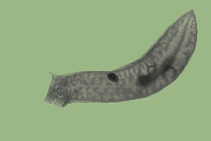
Figure 8. Mission accomplished. The Planaria glides away from the food pellets. Its body configuration has become shorter and wider; the pharynx, now internalized is visible in the distal part of the body. The gastrovascular cavity is also visible. There air bubbles inside the body. An ostracod is swimming on top of the Planaria body. The field is 1.1 cm wide.
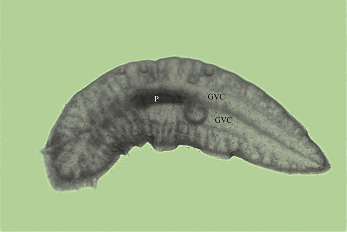
Figure 9. The Planaria at siesta. The body is maximally shortened, and the retraction of the chromophores allows visualization of internal organs such as the two branches of the gastrovascular cavity (GVC) and of the pharynx (P). In contrast, the eyes become near invisible. There are several air bubbles inside the body.
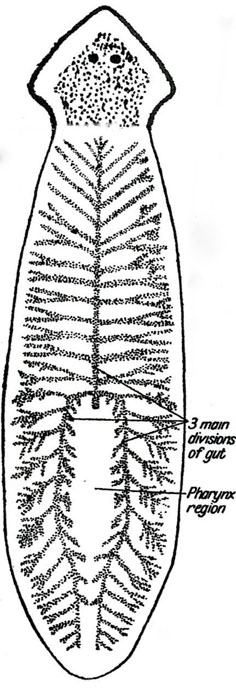
Figure 10. Diagram of the Planaria body identifying the digestive structures.
COMMENTS
• Planaria is an incredibly interesting animal. Evolution was displaying its full fancy when it developed it.
• We report a simple experiment carried on with elements well within reach of any amateur microscopist; it was both pleasurable to perform and informative in its results. If nothing else, it confirms the assertion made by somebody (Micscape, April 2007) that the micro-aquarium is: “A perennial source of amusement.”
• This is the ideal time of the year to gather your own supply of Planaria form almost any pond or slow stream. A water sample taken together with some bottom debris is likely to provide them.
Comments to the authors are welcome.
SOURCES
• Anonymous (2009) Planarian. http://en.wikipedia.org/wiki/Planarian
• Anonymous (2009) Slurpee. http://en.wikipedia.org/wiki/Slurpee
• Block, R.A, and J. V. McConnell (1967) Classically conditioned discrimination in the Planarian, Dugesia dorotocephala., Nature, 215: 1465-6.
• Buchsbaum, Ralph (1938) Animals Without Backbones. An Introduction to the Invertebrates. Ch. 10. The University of Chicago, Chicago, Ill, pp. 405.
[This is a classic book that went through numerous editions and re-printings and was offered in hard- and soft-cover versions. Even today it is a worthy item for the library of the amateur microscopist]
• Cooper, Joshep D. and Joseph C. Abbott (1979) Close-up Photography and Copying. Amphoto, Garden City, NY, p. 54.
• del Cerro, Manuel (2007) The Aquarium Microscope and The Micro-aquarium. A Perennial Source of Amusement. Micscape, April 2007
• Rilling, M. (1996). "The mystery of the vanished citations: James McConnell's forgotten 1960s quest for planarian learning, a biochemical engram, and celebrity." American Psychologist 51: 589–598. doi:10.1037/0003-066X.51.6.589
Published in the September 2009 edition of Micscape.
Please report any Web problems or offer general comments to theMicscape Editor .
Micscape is the on-line monthly magazine of the Microscopy UK web site atMicroscopy-UK
© Onview.net Ltd, Microscopy-UK, and all contributors 1995 onwards. All rights reserved.
Main site is at www.microscopy-uk.org.uk with full mirror at www.microscopy-uk.net .