|
Darkfield illumination with a desk LED lamp. By Rolf Vossen, Netherlands |
Introduction
Darkfield or darkground illumination is a very useful technique for visualizing specimens that are transparent and difficult to see in regular brightfield illumination. There are several ways to achieve darkfield illumination; the most frequently used methods include using opaque filter stops, phase annuli, or a specialized darkfield condenser. While creating darkfield at low magnifications is easy to accomplish, at medium to higher magnifications it can be a greater challenge. The method described here uses incident light from a desk LED lamp and no condenser at all to generate darkfield illumination at medium to high magnifications.
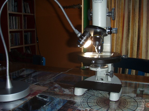
Fig.1: Setup with substage condenser removed to avoid reflections from the top lens.
Experimental setup.
Creating darkfield at low magnifications is usually not a problem so I restricted my choice of objectives to a 40/0.65 and a 60/0.85 achromat. I used a desk LED lamp that you can bend like a flexible fibre optic light guide from a cold light source and positioned it very close to the objective, illuminating the specimen from above/sideways (fig. 1). The lamp is called “JANSJÖ” (IKEA) and contains a 4.2 W LED that gives a bright “warm” light with a slight yellowish character. To get the darkest background you may have to play with the position of the lamp. Initially there were some lighter spots in the field of view and I quickly realized that these were due to light reflections coming from the condenser top lens. After removing the condenser the quality of the darkfield improved significantly. For making pictures I used a monocular microscope to get the most of the light from the specimen. When using a binocular microscope, the darkfield generated this way can be a little dim at higher magnifications and there may not be sufficient light for making photographs. I also put a piece of black plastic on the horse-shoe base of the stand to avoid any reflections from the table. Three different specimens were used to demonstrate the effect: a Klaus Kemp slide of Pleurosigma angulatum (fig. 2 and 3), cheek epithelial cells (fig. 4 and 5), and some filamentous algae (fig. 6 and 7). Pictures were taken through the eyepiece with a point and shoot digicam on a tripod stand. No image processing other than resizing was done after the pictures were taken.
Results
The darkfield created this way turned out to be of surprisingly good quality giving sufficient resolution and detail. The obvious difference with regular darkfield illumination is that the light is mainly coming from one direction giving it a more oblique character. With the 40x objective the dots of P. angulatum were easily resolved as they were with the 60x objective (fig 2 and 3). The pictures shown here however do not have the same clarity and details as seen directly through the eyepiece. I also tried low power objectives with good results.
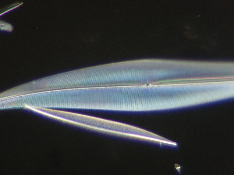
Fig.2: P. angulatum with 40/0.65 objective.
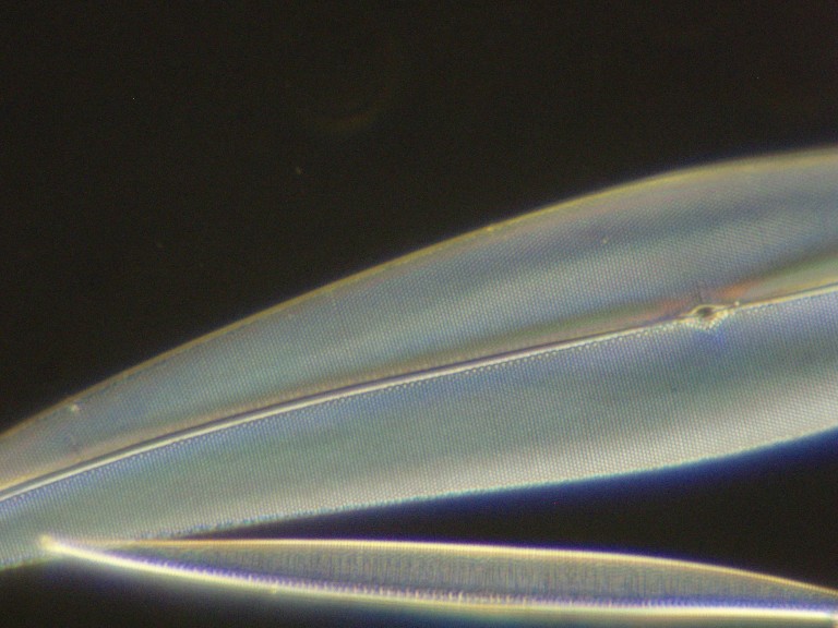
Fig.3: P. angulatum with 60/0.85 objective
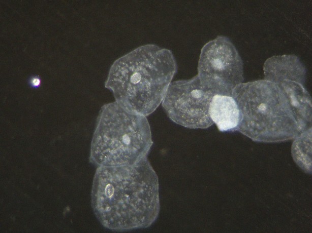
Fig.4: Cheek epithelial cells with 40/0.65 objective.
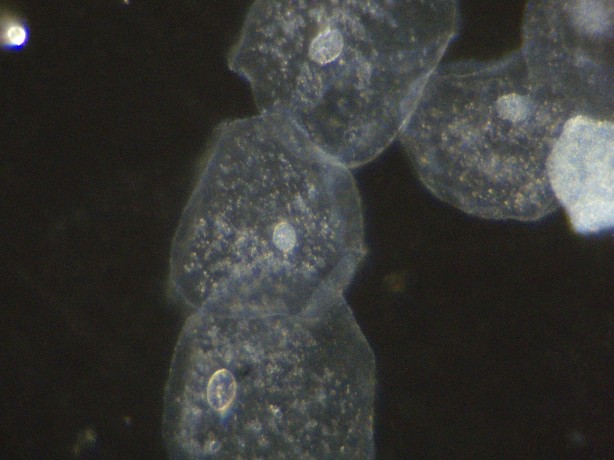
Fig.5: Cheek epithelial cells with 60/0.85 objective.
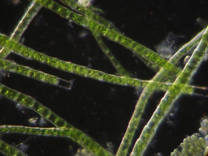
Fig.6: Filamentous algae with 40/0.65 objective.
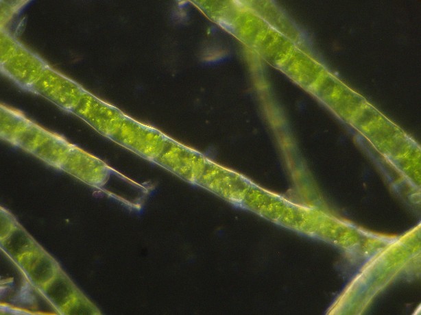
Fig.7: Filamentous algae with 60/0.85 objective.
Conclusion
Presented here is a very easy way to achieve darkfield illumination with a desk LED lamp and incident illumination. A stronger light source than the one used here may be needed when using a binocular microscope, otherwise the darkfield image can be dim at higher magnifications. Without the need for a condenser this method can be used on entry-level microscopes that often have no substage condenser with filter holder. While this method is by no means a replacement for a darkfield condenser it may be an attractive alternative to home-made darkfield stops.
Comments
will be welcomed by the author.
Microscopy UK Front
Page
Micscape
Magazine
Article
Library
Published in the September 2009 edition of Micscape Magazine.
Please report any Web problems or offer general comments to the Micscape Editor .
Micscape is the on-line monthly magazine of the Microscopy UK website at Microscopy-UK .
© Onview.net Ltd, Microscopy-UK, and all contributors 1995 onwards. All rights reserved. Main site is at www.microscopy-uk.org.uk.