The genus Campanula contains hundreds of
species, of which about ten are grown as houseplants. The
examples photographed in this article were obtained as
cut-flowers. Campanulas are commonly referred to as bellflowers
because of their bell-like shape. In fact, the name campanula
translates to bell. Some are long and tubular, while others are
shorter and more rotund. All of these flowers belong to the
family campanulaceae.
Although most family members are native to the Mediterranean and
mountainous Balkan countries, my examples were flown to Canada from a
greenhouse in Columbia. Air transport has certainly changed the
availability of flowers for household display! As you will see,
the stems contain flowers of different colours - pink, pale violet,
white, and white with pink lobes.
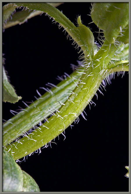
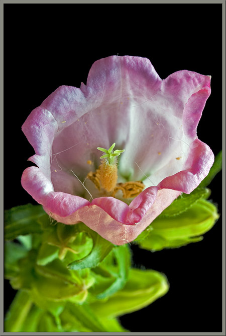
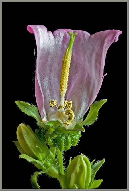
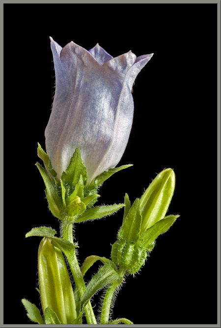
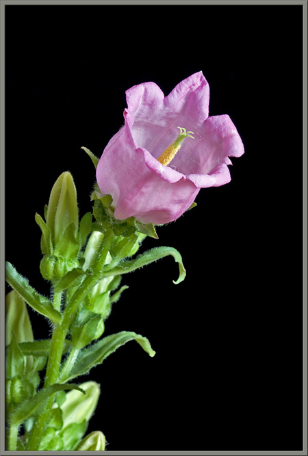
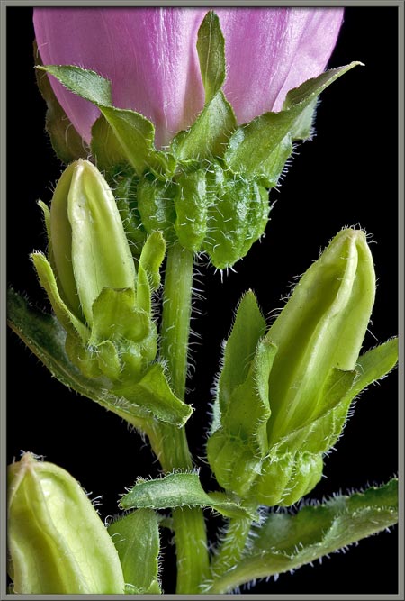
The image above shows the main characteristics of the bellflower.
A fused tubular corolla ends in six flared, pointed lobes. The
surfaces of stems, and the narrow leaves, are covered in fine
hairs. My samples had stems approximately 40 centimetres in
length.
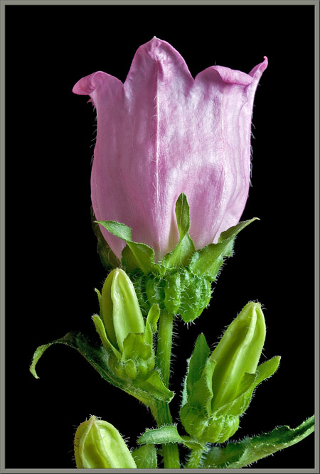
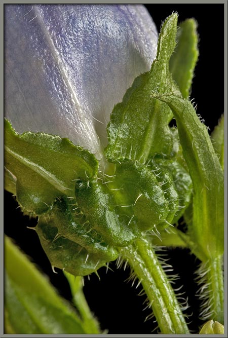
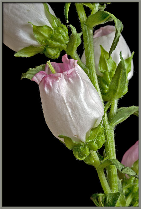
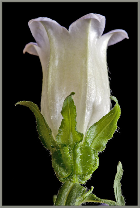
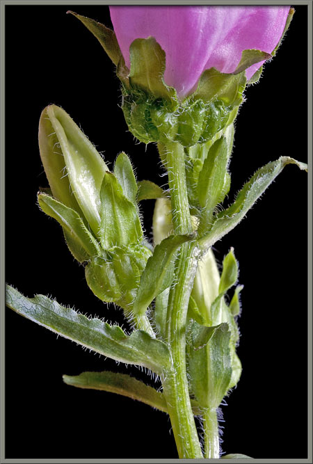
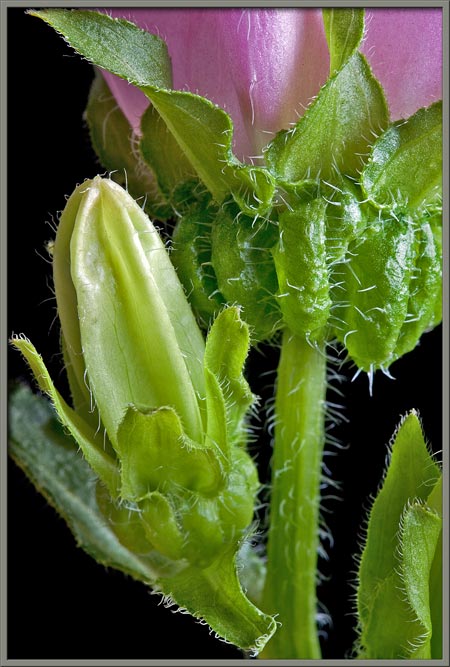
Beneath each flower and bud, there is an unusual bulbous,
pumpkin-shaped swelling. This is the ovary in which the flowers
seeds will develop. Note the short green bracts (modified
leaves), located above the swelling, that surround the base of each bud
and flower. Also notice that the early buds show no sign of the
flowers final colour.
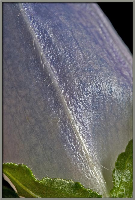
A close-up of the stem shows the many hairs growing from its surface.
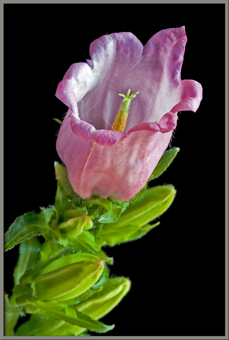
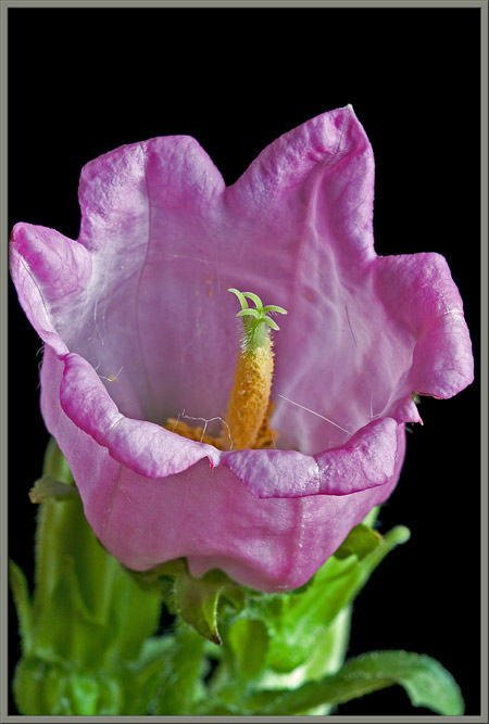
The images that follow reveal the overall shape of a mature
bellflower. If you look very closely at the corollas surface in
the right-hand image, you can see that it is covered in a myriad of
tiny downward pointing
hairs. These hairs discourage insects like ants from climbing to
the top of the flower. (Such insects are not efficient
pollinators, and this effectively prevents the possibility.)
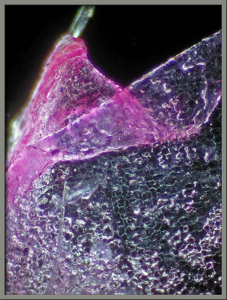
If the tip of one of the corolla lobes is examined under the
microscope, its cellular structure is visible.
When the corolla is viewed from the front, the flowers reproductive
structures can be seen. Two additional adaptations to prevent the
entry of small insects into the flowers mouth are also visible.
Note that the corolla lobes curl backwards, forming a barrier to
climbing insects. If you have good visual acuity, you may also be
able to see a number of long hairs that grow from the top of the
corolla and crisscross the flowers mouth. These too help
discourage the entry of small insects. Large ones, of course,
have no trouble pushing them aside.
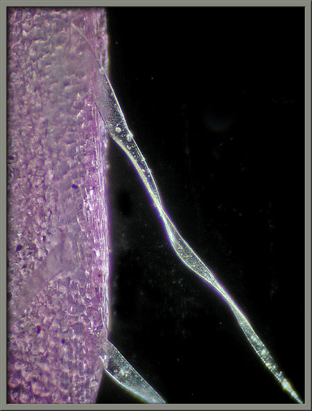
The photomicrograph below shows a couple of these long hairs and their
points of connection to the corolla.
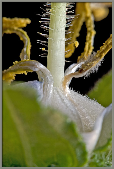
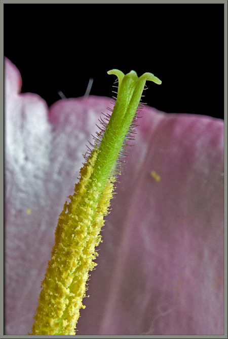
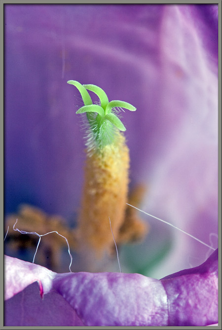
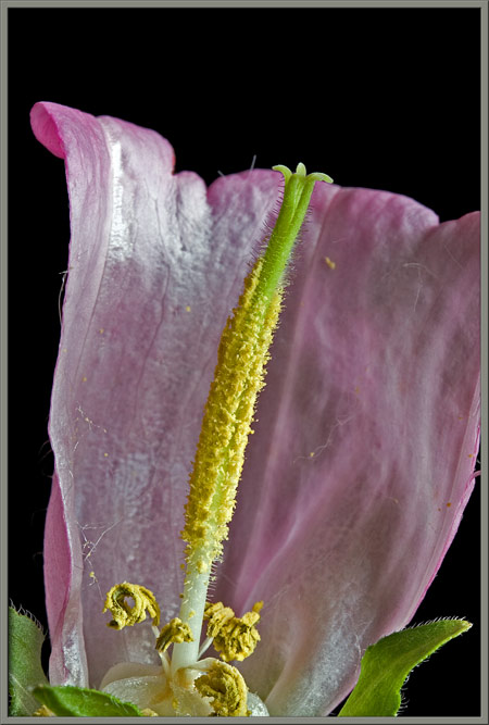
Cutting away a section of the corolla allows the reproductive
structures to be seen more clearly. The flowers pistil is composed of
a long pollen encrusted style which supports a lobed stigma (the female
pollen accepting organ). Clustered at the base of the style are a
number of curled stamens with their yellow anthers (male pollen
producing organs) supported by short filaments.
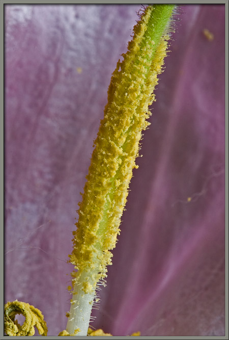
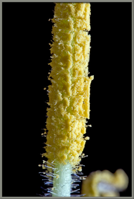
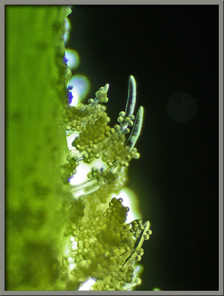
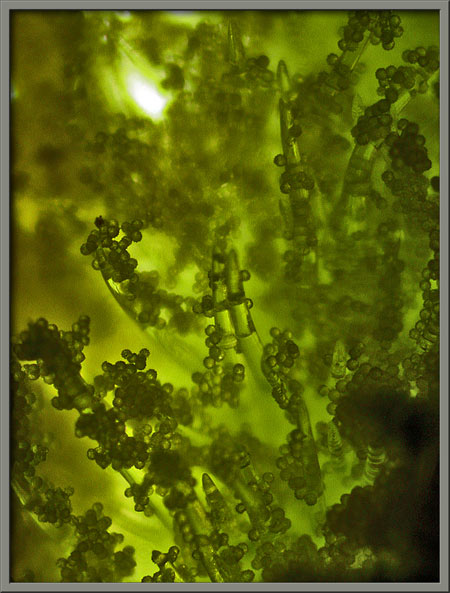
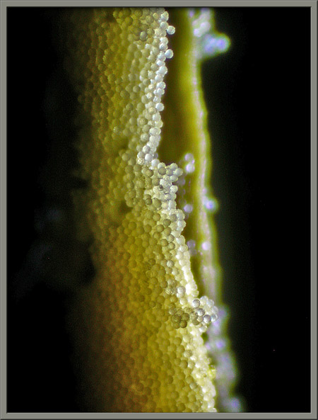
A close-up of the style reveals that it is covered with hairs which
retain copious quantities of pollen.
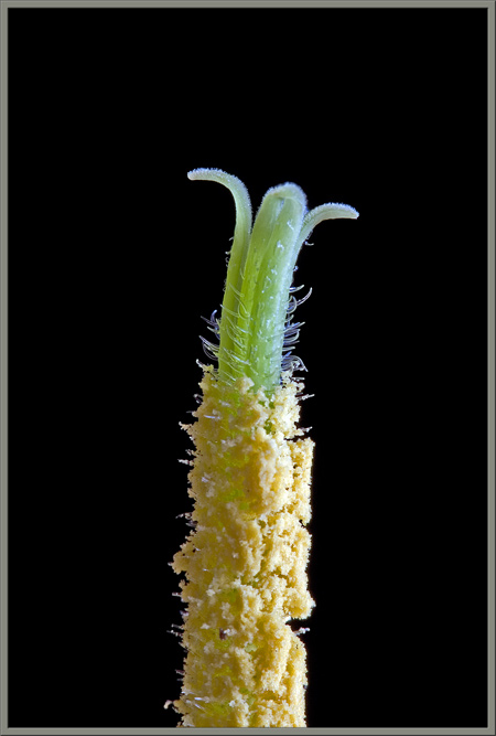
An even higher magnification of the base of the style shows these hairs
more clearly.
The microscope provides an even better view of the hairs and the
spherical pollen grains that cling to them.
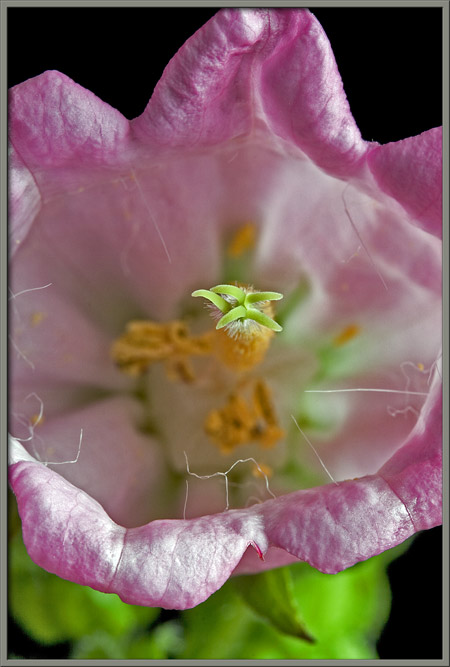
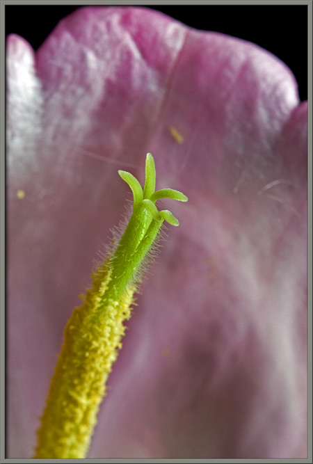
The images that follow show the stigma itself. Notice that it is
composed of five rather long lobes which curl back to be perpendicular
to the stigmas base. (The third image provides another look at
the long hairs at the corollas edge.)
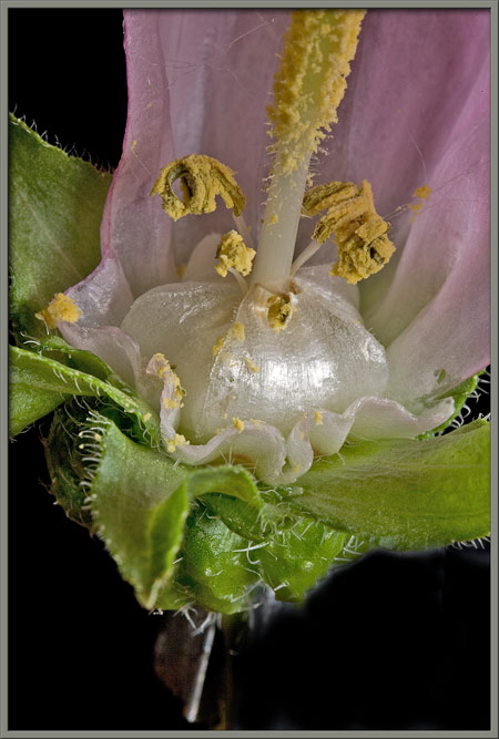
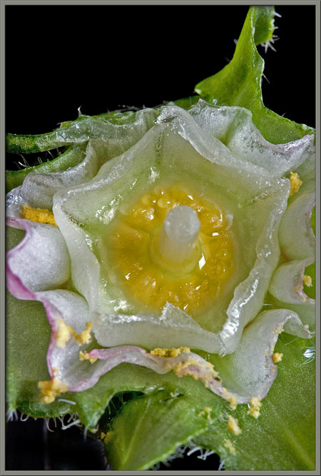
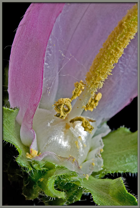
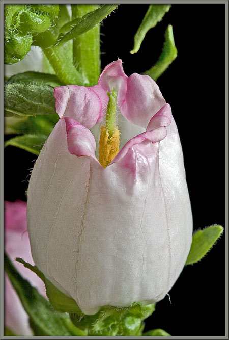
The bellflower seems to erect many barriers to prevent insects from
obtaining the flowers nectar. Here is another one. At the base
of the corolla, the pool of nectar seen in the image at right is
covered by a number of curled, white, petal-like structures that form a
protective dome. In order to get at the nectar, the insects
proboscis must pass between the petal segments. In doing so the
insect (most likely a bee), must be close enough to brush against the
anthers that are positioned just above the dome. How amazing are the
schemes that the plant kingdom has evolved in order to facilitate
fertilization!
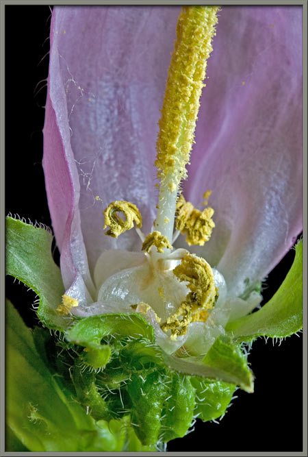
Now lets look more closely at these stamens. As can be seen in
the three images below, the yellow anthers are tightly coiled, and held
in position by relatively short white filaments. The bellflower is
protandrous; the stamens become mature before the stigma. This
helps prevent self-fertilization.
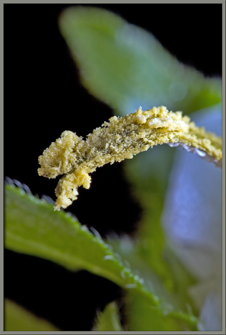
A close-up shows the large number of pollen grains clumped on the
anthers surface.
Under the microscope, the under-surface of the stigma appears to be
relatively smooth, while the upper surface is covered with the
hair-like protuberances that can be seen in the right-hand image.
Several photomicrographs follow that show the accumulation of pollen
grains on an anthers surface.
**********
Some years after this article had been written, Samuel Jordan, a botanist
with much greater knowledge about the plant than I have, sent me the following
information. I very much appreciate his willingness to share his knowledge with
me and my readers.
In my search of information about nectar in bellflowers I stumbled across
your article. The pictures are amazing and it is very informative. But might I
point out that you are not quite correct about how insects pick up pollen. As
far as I know, it happens as follows:
-The anthers open when the flower is still closed. As they are appressed to
the style, most of the pollen gets caught in the hairs covering the style.
-The flower then opens. When the flower opens the anthers are already
wilted and the pollen is caught in the stylar hair.
-Insects then come to collect nectar, which is hidden behind these, as you
call them, "petal-like structures" at the back of the flower. Doing so they rub
against the style covered in pollen and pick some up.
-Mechanical rubbing of insects against the pistil stimulates the retraction
of the hairs, so that when the next insect comes, there is again some very loose
pollen that will easily fall off. This goes on until all the hairs have
retracted and there is no more pollen left on the style.
-The stigma then open and are ready to pick up pollen from visiting insects
(the stigma can sometimes open before all hairs have retracted).
**********
Here is the stem containing pale violet flowers that was mentioned earlier.
Closer views of the flowers base, and detail in the corolla tube
itself, can be seen below.
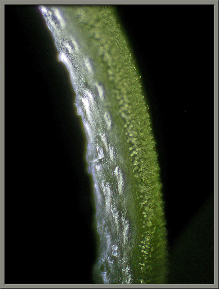
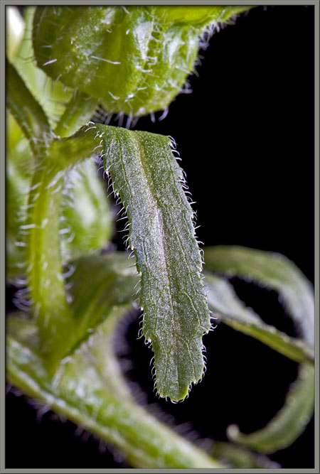
Notice the hairy edge of one of the plants leaves.
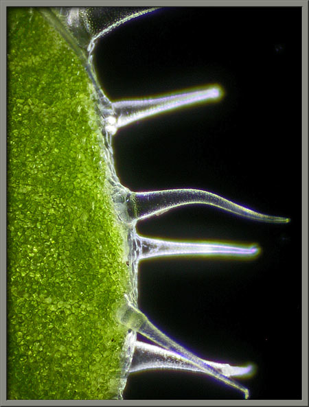
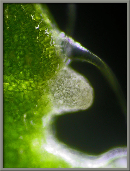
If the edge of one of the bracts immediately below the base of the
corolla is examined under the microscope, hairs are clearly visible.
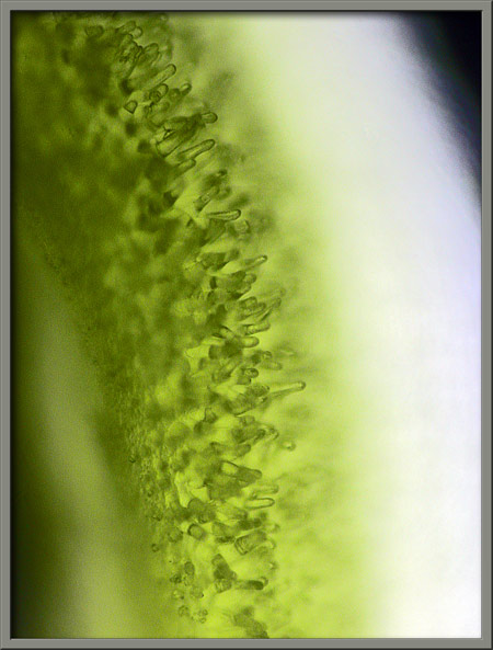
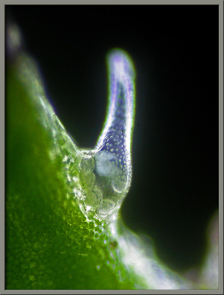
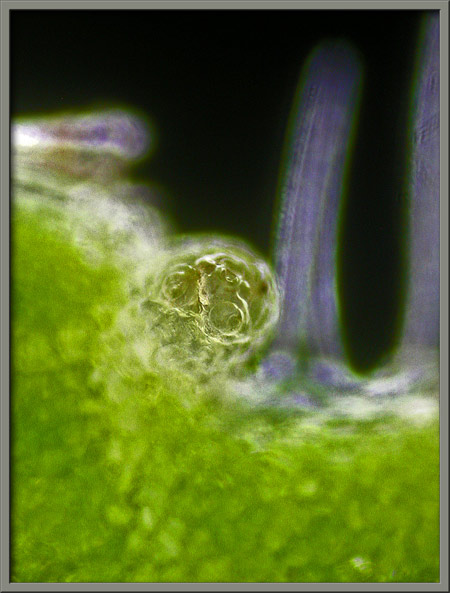
Higher magnification reveals still more details. Notice that the
base of each hair is ringed by light green spherical cells. Also
note the circular dimples that cover each hairs surface.
Adjacent to each hair is a small lumpy protuberance. (I have been
unable to determine the function.)
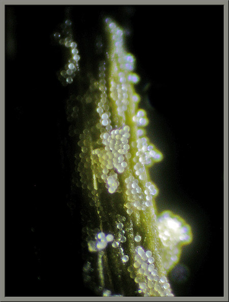
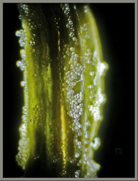
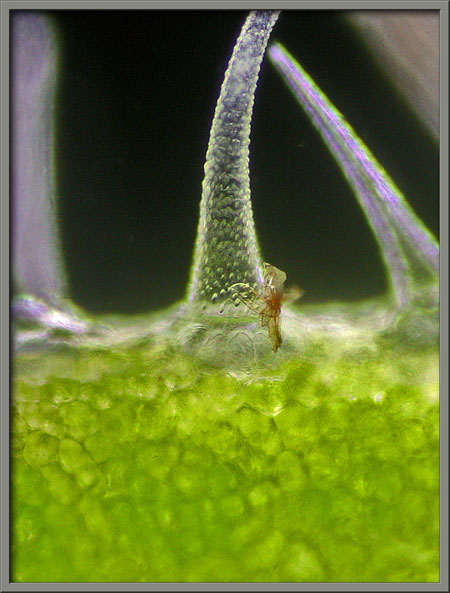
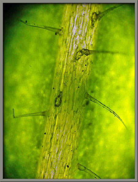
The photomicrograph below show hairs growing from the central rib of
the bract. The black specks are carbon soot. (Amazing -
Columbia must have air pollution!)
Finally, here are several images showing the colouration of other
bellflowers.
Photographic Equipment
The macro-photographs were taken with an eight megapixel Canon 20D DSLR
equipped with a Canon EF 100 mm f 2.8 Macro lens which focuses to
1:1. A Canon 250D achromatic close-up lens was used to obtain
higher magnifications in several images.
The photomicrographs were taken with a Leitz SM-Pol microscope (using
dark ground and phase-contrast condensers), and the Coolpix 4500.
A Flower Garden of
Macroscopic Delights
A complete graphical index of all
of my flower articles can be found here.
The Colourful World of
Chemical Crystals
A complete graphical index of all
of my crystal articles can be found here.