Beyond the Diffraction Limit – Pond Dipping with an Electron microscope
by Ian Portman, UK
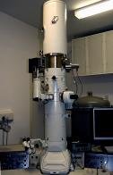
I have many microscopes at home but none are quite as nice as the microscopes I have at work. I’m an electron microscopist and this is my microscope –
It’s a Jeol 2010f field emission transmission electron microscope it has a top magnification of 1 million x, normally it’s used for looking at individual protein molecules but what started as a flippant comment about pond dipping has resulted in few skipped lunchtimes and some nice photographs.
Staining – As with many light microscope specimens, TEM samples need staining. Often we use uranium acetate, it’s electron dense so creates good contrast in the microscope and it’s cheap to buy. Though often called a negative stain it reacts with specimens in complex manner and can produce positive staining effects.
Our local ‘pond’ has dried to little more than a shallow mud puddle, even the ducks have deserted but there’s still life to be found. A quick filter gets rid of the bits of mud, water boatmen and ubiquitous daphnia that are too big to get under an electron microscope. Then a low speed centrifuge, to pull down all the larger cells –
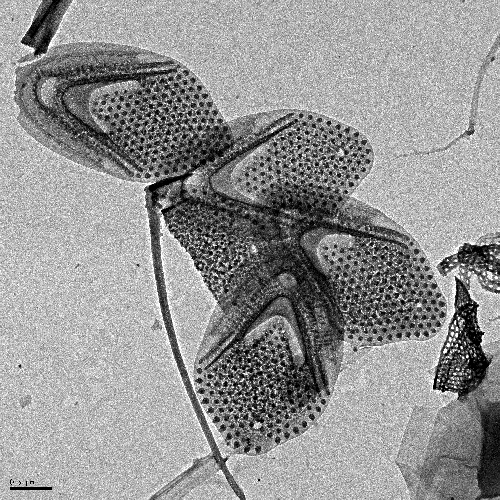
Possibly diatoms(?) at 8000x. Stained with uranium acetate.
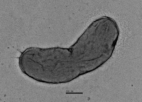
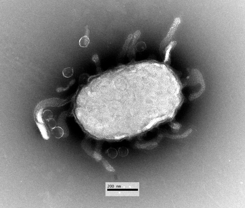
A Bacterial Cell, the halo around the right hand end is caused by pili – hairlike appendages on the surface of the cell. Just visible on the left hand side are the flagella that the bacterium uses to swim.
The apparently clear water left over still contains many things, collecting these needs another centrifuge, this time running far faster at 40,000rpm – enough to spin down things a fraction of a micron across.
Now we’re at the scale I normally work at. With all those bacteria in the pond, there must be their parasites in abundance – bacteriophages, viruses that live off bacteria.
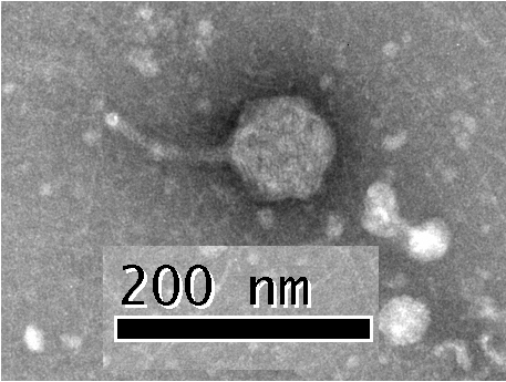
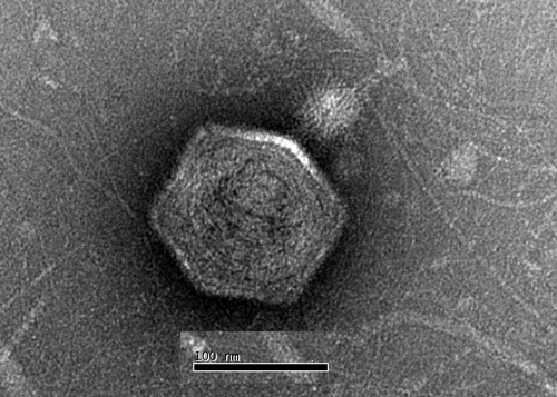
A typical bacteriophage with a head, tail and tail fibres.
An infected cell buds out fresh viruses, within the cell new virus heads can been seen forming.
Bacteriophage come in different shapes and sizes, this species is short tailed.
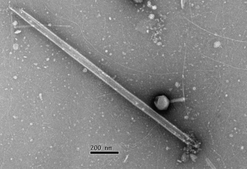
A less typical form of phage, these filamentous forms carry their DNA in a linear form and can get to a micron in length.
The Mystery gallery
I’m open to suggestions on what this object might be.
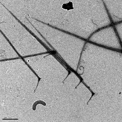
Filamentous objects at 6000x- algal cells perhaps?
All images were taken in the University of Warwick School of Life Sciences Imaging suite.
Comments to the author will be welcomed.
Microscopy UK Front
Page
Micscape
Magazine
Article
Library
Published in the September 2010 edition of Micscape Magazine.
Please report any Web problems or offer general comments to the Micscape Editor .
Micscape is the on-line monthly magazine of the Microscopy UK website at Microscopy-UK .
© Onview.net Ltd, Microscopy-UK, and all contributors 1995 onwards. All rights reserved. Main site is at www.microscopy-uk.org.uk .