
|
Another Marine Mystery: A Sea Cucumber With Jaws??? by Richard L. Howey, Wyoming, USA |
Well, O.K., not really jaws, but odd little structures called pedicellariae which are generally associated with starfish (asteroids) and sea urchins (echinoids), but can we trust Mother Nature? However, before we proceed, let’s briefly consider the nature of pedicellariae which are, as a matter of fact, extraordinarily curious structures.
There are a number of different types and the primary function which they serve is to discourage or eliminate “settlers”; that is, larvae and small organisms which have an opportunity to settle on the surface of a sea urchin or starfish and take up residence. When the pedicellariae are triggered, they can crush a larva or small organism by means of their “jaws”. The two basic forms of pedicellariae are stalked or sessile; the former have a calcareous rod covered by a membrane with the jaws at the top of the rod whereas the sessile forms develop directly on the surface of the starfish or sea urchin.
The sessile forms usually consist of just 2 or 3 jaws; these are sometimes found in clusters ringing the base of the spines or on a few species they develop directly on the sides of the spines. The types with only 2 jaws are valvate and vaguely like a bivalve mollusk in appearance except that both ends are open.
Among the stalked pedicellariae there is a considerable degree of variation; some have only 2 jaws, but others have 3, 4, or even 5. Among the 2-jawed ones are the “forceps” and “scissor” types. There are some specialized pedicellariae that even have minute poison glands and there are some elaborate and quite formidable-looking types with crossover-toothed jaws that catch small fish and there is even a form that has clusters of pedicellariae which when agitated can rise on a stalk, like an hydraulic lift that a garage mechanic would use. As I said earlier, these structures are mostly defensive, but some actually capture prey, apparently digest it, and function almost like an independent organism or part of a colony. So, as you can gather they are extraordinarily interesting structures.
However, sea cucumbers aren’t supposed to have pedicellariae! So, what do we make of this specimen which I have? Let’s begin with what you need to know about the specimen (and, of course, I have only one of this particular species, so I can’t compare it.)
1) It is from the Philippines and was shipped with about 20 other specimens of sea cucumber, but no starfish or sea urchins. However, before they were shipped, they might have been consorting with another order of echinoderms, who knows? My examination of my victim began, as it frequently does, by taking scrapings from the surface with a surgical scalpel and placing these in a drop of water on a slide. Using my stereo-microscope, I could see that I had some bits of surface tissue and some fragments of tube feet.
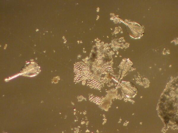
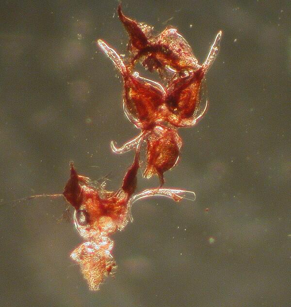
2) The type of pedicellariae I am finding here are tridentate, that is, they have 3 jaws as you can see from the image below, even though they are no longer attached at the base.
3) At this moment, the question of whether these are sessile or stalked is still unresolved independently of the issue of whether or not these structures genuinely belong to this sea cucumber or not.
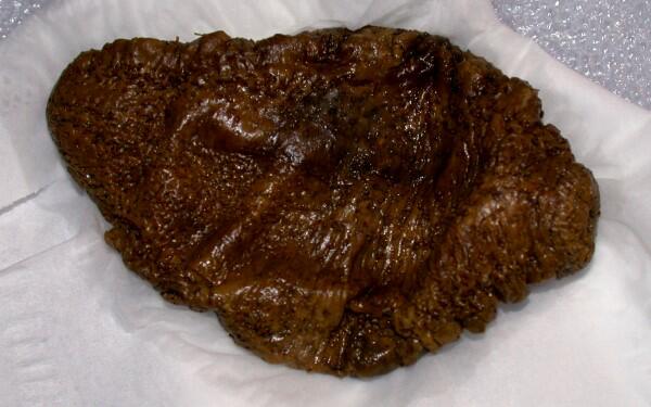
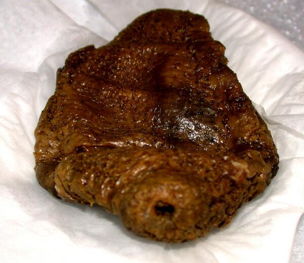
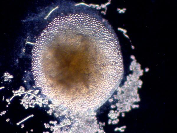
4) However, I have been remiss in that I haven’t shown you what the organism itself looks like. It certainly isn’t distinguished; it has a rather typical form somewhat like a small Cucumaria, blandly brown and, in this case, quite flattened. At the anterior end, one can see the oral opening through which the tentacles project when the organism is alive.
5) When taking scrapings and when treating them with household bleach (sodium hypochlorite), I noticed fragments of the tube feet. I observed elaborately fenestrated plates and now I am curious as to whether or not these are broken fragments of a more connected structure within the tube feet or whether these are simply typical.
Now what I am going to do is isolate some tube feet, place them in tubes, subject them to bleach and see what I get. I will also take a more or less intact tube foot or two, place them on a slide and add bleach and then carefully observe them in stages as the tissue is disassociated from the calcareous structures. I learned quite some time ago that the tube method can produce misleading results as a consequence of radical disassociation of the component parts.
I isolated 2 tube feet and then, using a micro-scalpel, cut off just the very tip of the terminal end. I added some bleach and then watched carefully as the tissue began dissolving away.
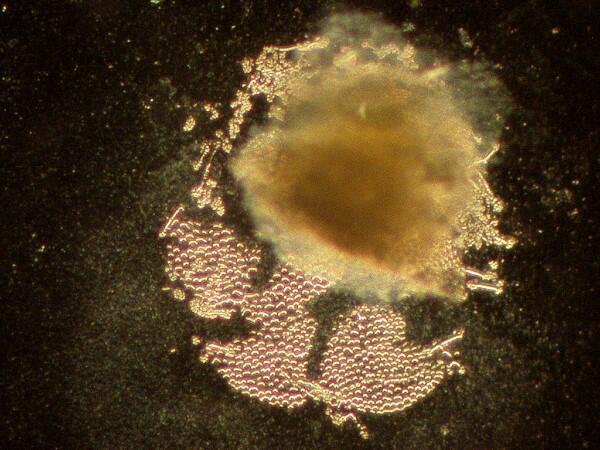
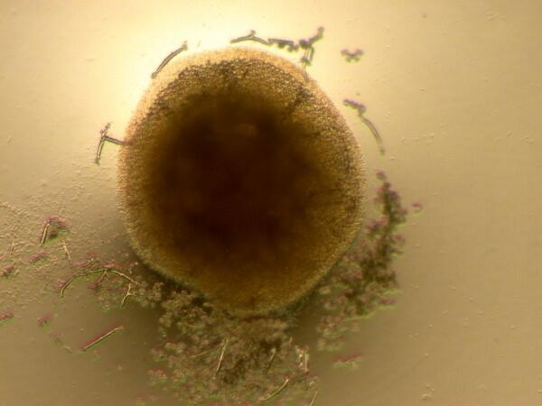
Using one of my micro-fine needles made with a Minute-Nadel insect pin, I would, every minute or so, very carefully move away the raft of air bubbles forming over the tube foot tip as the tissue dissolved in an effort to get an unobstructed glimpse of the surface. After several times, there emerged, to my delight, a round fenestrated disk under which I am fairly sure there is a second such disk, but demonstrating that will require further investigation.
The surface tissue in the podia (tube feet) of this species is translucent and when I examine longer segments of the podia, I can see milky-white calcareous material at several locations under the surface of the epidermis. Now, the question becomes an issue of whether or not these are also disks or just rather randomly-shaped fragments which form within the podia? The next question has to do with the function of these structures.
When I dissolve out the tissue, I do indeed find calcareous fragments rather than coherent disks like those in the tips of the podia.
This suggests that these are not support structures, so their possible function remains unclear. (Possibly just another one of Mother Nature’s games to lead us down the garden path.) Here again, we need to remind ourselves that the colossal number of random experiments which occur in the small parts of the universe that we are partially familiar with, are so vast that it may well be that these structures once had a definite, concrete function, but have become vestigial or maybe we just haven’t looked and thought carefully enough to discover their purpose or perhaps they are simply a consequence of cosmic playfulness and whimsy. That, of course, would be an outrage, because science is supposed to be rational methodical, sensible, and orderly (except, I guess, when it comes to quantum physics.)
For many years, the physical sciences dominated, but now we are gradually beginning to understand that the challenges which the biological sciences present to us are equal to or surpass those of the physical sciences. (As a brief aside, just think how much it costs to do particle/quantum physics–billions for the Hadron Super Collider. Imagine the kinds of intricate technologies that could be brought to bear on trying to understand the extraordinary organisms with which we share this planet if that kind of money were available. Some years ago, the price of one nuclear submarine (without cost overruns) was estimated at 2.6 billion dollars and, of course, the Department of Defense couldn’t settle for just having one. However, think what the price of just one could make possible in terms of producing a number of deep-sea submersibles that would make Alvin seem like a Model-T Ford. We, as human beings, need to start seriously reconsidering our priorities in terms of the allocation of resources. O.K., end of rant–back to the sea cucumber. However, one more comment relating to quantum physics; it suggests that there are certain things (states of affairs) that are intrinsically uncertain and thus unknowable. I certainly think that this is true in relation to biological systems, but this does not provide a good methodological, procedural basis and so, stubborn empiricist that I am, I keep looking for explanations.
Once I discovered that the pedicellariae are to be found in small clumps on the surface and especially around the podia, both at the top and around the base, as well as in the relatively deep wrinkles in the skin of the sea cucumber, I knew where to hunt for them. I have continued to look for any hints of rods or stalks that might indicate that these pedicellariae do indeed belong to this organism. At one point, I was briefly encouraged (about 10 seconds worth) when I noticed that in several instances, the pedicellariae seemed directly and tenaciously attached to the very tips of some of the podia. However, further examination quickly revealed that what had occurred was that a very sharp tooth of one of two of the jaws had become embedded in the tissue of the terminus of the tube foot. (Not too bad for alliteration.) This type of sea cucumber crawls along the bottom feeding on the detritus of the substrate. So, if echinoids (sea urchins) and asteroids (starfish) are the only 2 groups of echinoderms which possess pedicellariae, then the puzzle of the presence of the pedicellariae here persists. (Oh, I really think I should receive the Micscape Monumentally Momentous Monotonous Meritorious Medal for alliteration.)
It is very difficult for me to imagine this rather sedentary creature suddenly launching an attack on a sea urchin or starfish and thereafter proudly wearing the pedicellariae as ribbons of honor.
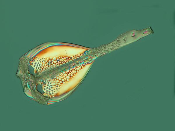
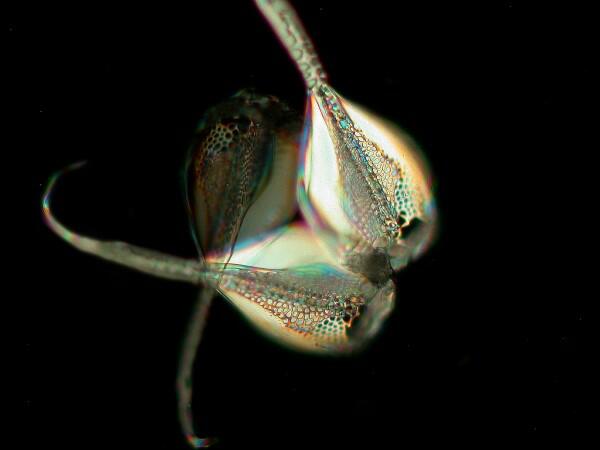
These pedicellariae are tridentate and the individual jaws are rather like an elongated bowl of a spoon, but at the tip form a narrow point with a curved, very sharp hook.
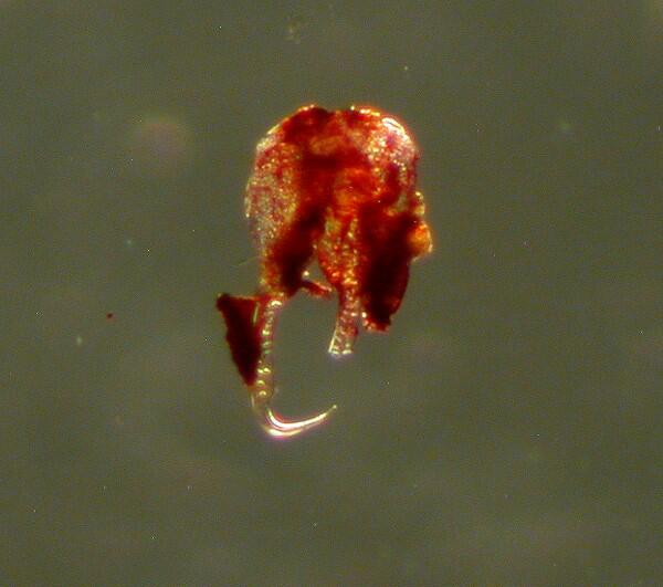
These are difficult to photograph for several reasons: 1) They do have a covering of tissue which, as you notice, is, in this case, a deep red color. 2) They tend to occur in clumps with the jaws open and sometimes interlocked. 3) When disassociated using bleach, the process is not controllable and, as a consequence, the result is separation of the jaws. 4) When the jaw is lying on the substrate, the sharp hooked tooth is virtually impossible to see. Getting a side view which reveals the tooth is usually possible only with a broken fragment as in the image below.
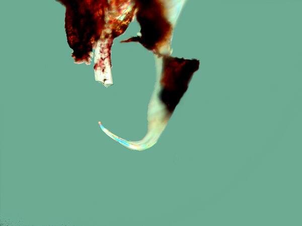
As you can see, these teeth are sharp, curved, and formidable.
5) Even the individual jaws, which because of their shape, I refer to as “mandolins”, are thick enough that it is not really possible to get an image which is fully in focus to show its detail. As a consequence, one has to use that brilliantly, marvelous invention called “stacking software”. This involves taking a series of images at different focal planes. I start at the very lowest point where that lowest bit is just barely in focus, take an image and then focus up just a couple of microns, take another image, and repeat until I get to a top point where that part of the specimen is losing focus. For this particular pedicellariae jaw, I took 32 images with Nomarski and 27 images for picture with polarized light. Here’s where the magic begins; I download the images to my computer, activate the Helicon Focus software and it takes the 32 Nomarski images and compiles them into a single image and then with some further instructions takes the 27 polarized images and compiles then into a separate single image. Then I take my old graphic software program PhotoImpact XP–which isn’t even the most recent version, but I’m comfortable with it and I like it–and make a few refinements to the image. I realize that most tech-savvy people these days use Photoshop or Photoshop Elements. Hey I’m 173 years old and I found them overly complicated, not very “user-friendly”, and full of stuff that I don’t need and don’t want to learn. Remember I’m an old fossil–Professorus Emeritus Rex. If you can convince me that it’s really terrific, and I need it, then I’ll hire a university freshman who’s majoring in Computer Science.
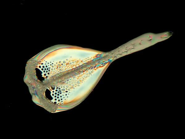
In any case, here are my paltry, antediluvian results. First the Nomarski images. I’m going to show you 3 of the same view to justify all the time I spent playing around with the computer
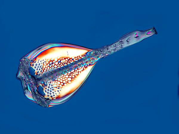
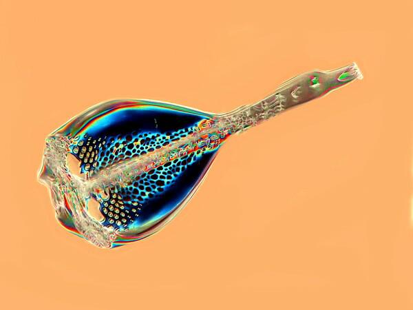
And now here is an image using polarized light.
At the tip of the “mandolin” is where the curved, hooked tooth is located but, because of the angle, it’s not visible in the images above, so here are 2 views of a small cluster of “mandolins” where the teeth are quite evident.
This afternoon, I decided to do some additional poking and scraping where there are pedicellariae. Once again, I was struck by the fact that there are well over 100 of them scattered around–perhaps more than 200–and also by the fact they tend to be located on or at the base of the tube feet or else in the relatively deep crevasses in the surface. This latter point is difficult to assess the significance of, since the sea cucumber in its living state would be much more rounded, “inflated” as it were, so these declivities in the surface would be less deep. I mentioned previously that I was also struck by the number of pedicellariae that had one or more of its teeth embedded in the tip of “stalk” of a tube foot and sometimes in surface tissue.
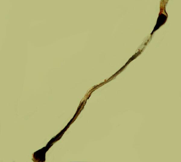
Today, I also found something which heightens the mystery. I found an example which has a rather long stalk, at least several times the length of the jaw. I had noticed some “thread-like” structures before but, I dismissed them because it was not clear that they were confected to the “head”, that is, the jaws. If the stalk’s base was attached to the sea cucumber that was not discernible. I did, however, slowly and carefully, using some delicate micro-tools manage to remove the jaws with the stalk still attached and isolate it in a tiny dish containing distilled water.
(And yes, a separate article on micro-tools is still forthcoming; I’ve just been too slothful to get around to taking the photographs.)
From my very cursory examination, ths structure doesn’t seem to possess calcareous support material. It is the case that not all such stalks have supporting rods (which would, of course, increase their flexibility). On the other hand, after what this specimen has been through, it would not at all be surprising that if it had possessed any calcareous supports, they might well have dissolved away. In any case, what is now required is a careful examination of the stalk with a compound microscope.
As you will see from the images, there is still some pigment tissue associated with the jaws. The tissue of the stalk has bent away from the jaws; however, within this narrow sheath of tissue there is clearly a long structure with a “knob” at each end. The question now is whether or not this is a calcareous supporting rod typical of certain forms of pedicellariae. As it turns out, the answer, “Callooh! Callay!” turns out to be an emphatic “Yes” as can be seen from the images below. Here is a low power image taken with my stereo-dissecting microscope
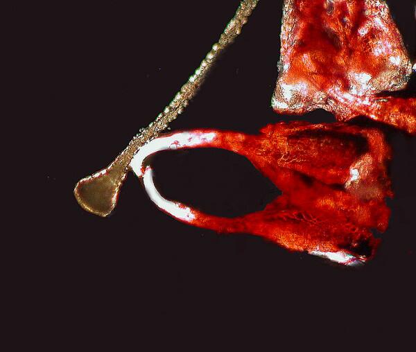
This is indeed provocative, because it is the first concrete hint I have been able to find of a calcareous support rod. Since this was removed directly from the sea cucumber in alcohol, transferred into distilled water, and not subjected to any other chemical treatment, I transferred it to a slide, using a micro-pipet to remove the water and then added a drop of high viscosity Type B immersion oil and a cover glass. I took the a few preliminary pictures with brightfield and Nomarski but, my primary interest was in obtaining some images with polarization to determine whether or not this structure is birefringent. It turns out that indeed it is and here is an image.
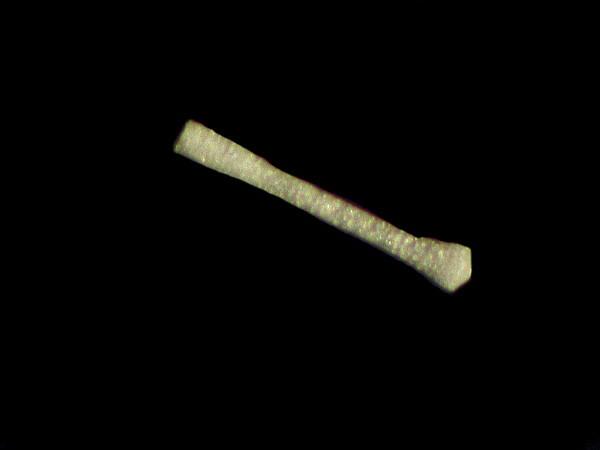
One end of the rod has less tissue and the “knob” there shows up in a quite clear fashion. As I examined the rod moving the lens down toward the other end, I noticed that as I approached the other “knob”, there was an area where the rod had broken and fragments of the calcareous crystals of which it is composed were visible.
I spent a couple of hours scanning the surface of the sea cucumber hoping to find additional examples of stalks with supporting rods but, with no success (yet).
Clearly, what I would most like to find are stalks that are unquestionably anchored, genuinely attached to the surface tissue of the sea cucumber itself. Right now, this seems unlikely.
However, suppose for a moment that this is a rare sea cucumber that actually does have pedicellariae, a basic issue would still remain: Why are there so many instances where the teeth of the pedicellariae are hooked into the tube feet or their tips? One possible answer is that a strange set of conditions triggered a “self-attack”. In other words, when the sea cucumber was removed from its natural environment and subjected either to drying or immersion in a preserving fluid, the pedicellariae responded to this as an attack and the only objects it could find that constituted a threat were the tube feet or surrounding tissue. I admit that this is an unlikely scenario but, nature is full of oddities, puzzles and anomalies so, for now, I have adopted a neutral position; perhaps out there somewhere in the vastness of the ocean realm, there are some sea cucumbers with pedicellariae.
It is these hooks or “teeth” that I have found embedded in the tips of tube feet, in the basal area of the tube feet, and in the surface tissue not directly associated with tube feet that raise the most serious doubts. From my relative casual examinations thus far, based on the general morphology of these pedicellariae, I would hazard the guess that, if they don’t belong to the sea cucumber (which they probably don’t) then they are more likely to come from a sea urchin rather than a starfish.
It is known that some types of sea cucumbers go dormant for weeks or even months at a time, so we might imagine this specimen hibernating at a spot down on the bottom when a sea urchin dies or is killed by a predator and the remains come drifting down and settle on the sea cucumber. Two immediate flaws regarding this scenario immediately pop into mind: 1) A large majority of echinoids are also bottom feeders and, as a consequence, are highly unlikely to become a rain of debris and 2) if such an event were to have occurred, then there surely would also have been some small (or even medium-sized) spines that would also have landed on the surface and ended up in the surface folds of the tissue of the sea cucumber. so , we really do have a singularly odd puzzle.
I admit that with this particular specimen, I have still just scratched (or scraped) the surface. I will continue to poke, prod, section, and dissect, and most of all, look for structures which suggest that the pedicellariae really do arise from and are attached to this exasperating sea cucumber. I’ll keep you informed.
All comments to the author Richard Howey are welcomed.
Editor's note: Visit Richard Howey's new website at http://rhowey.googlepages.com/home where he plans to share aspects of his wide interests.
Microscopy UK Front
Page
Micscape
Magazine
Article
Library
Please report any Web problems or offer general comments to the Micscape Editor .
Micscape is the on-line monthly magazine of the Microscopy UK website at Microscopy-UK .