Symbion pandora- revisited
by Mol Smith, UK
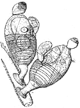
Way back in time when Micscape Magazine and this holding web site (www.microscopy-uk.org.uk) were both getting started, a new life form was discovered and reported on by us...
"Just before Christmas 1995, a
new life form discovered by Danish biologists on the lips of the Norway
lobster attracted world-wide media attention. Why? This topical article
by Dave Walker describes and illustrates this "zoological highlight of
the decade" using the biologists' published material and includes an exclusive
photograph kindly supplied by the original researchers".
{Read the article here}
We thought it was time to revisit Symbion pandora to see what information has come forth since that first pre-1995.
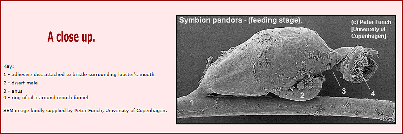
So let's look a little closer first via this SEM image from Peter Funch
Microanatomy and Development of the Dwarf Male of Symbion pandora (Phylum Cycliophora): New Insights from Ultrastructural Investigation Based on Serial Section Electron Microscopy.
Ricardo Cardoso Neves and Heinrich Reichert
(2015), PLoS ONE. Abstract from paper quoted below.
"Cycliophorans have a complex life cycle that involves several sexual and asexual stages. One of the sexual stages is the 40 μm-long dwarf male, which is among the smallest free-living metazoans. Although the dwarf male has a highly complex body plan, this minute organism is composed of a very low number of somatic cells (~50). The developmental processes that give rise to this unique phenotype are largely unknown. Here we use high resolution serial block face—scanning
electron microscopy to analyze the anatomy and morphogenesis of three cycliophoran dwarf males at different developmental stages ranging from internal bud to mature male. The anatomical and morphological features of the mature dwarf male stage reported here largely correspond to those reported in earlier studies. Interestingly, the organs that typically characterize the anatomy of the mature dwarf male, e.g., muscles, brain, testis and glands, are already formed in the young male. However, there are striking
differences between the mature male and young male stages at the level of cellular architecture. Thus, while the young male stage, like the internal bud stage, possesses approximately 200 nucleated cells, the mature male stage comprises only around 50 nucleated cells; muscle and epidermal cells of the mature male lack nuclei. Moreover, the total body volume of the mature male is only 63% of the body of the young male implying that the maturation of the young male into a mature male involves a marked reduction
of internal body volume, mainly by massive nuclei loss. Our comparative analysis of these dwarf male specimens reveals unprecedented insight into the striking morphological and developmental differences that characterize these highly miniaturized male stages both at the level of body organization and at the level of cellular ultrastructure."
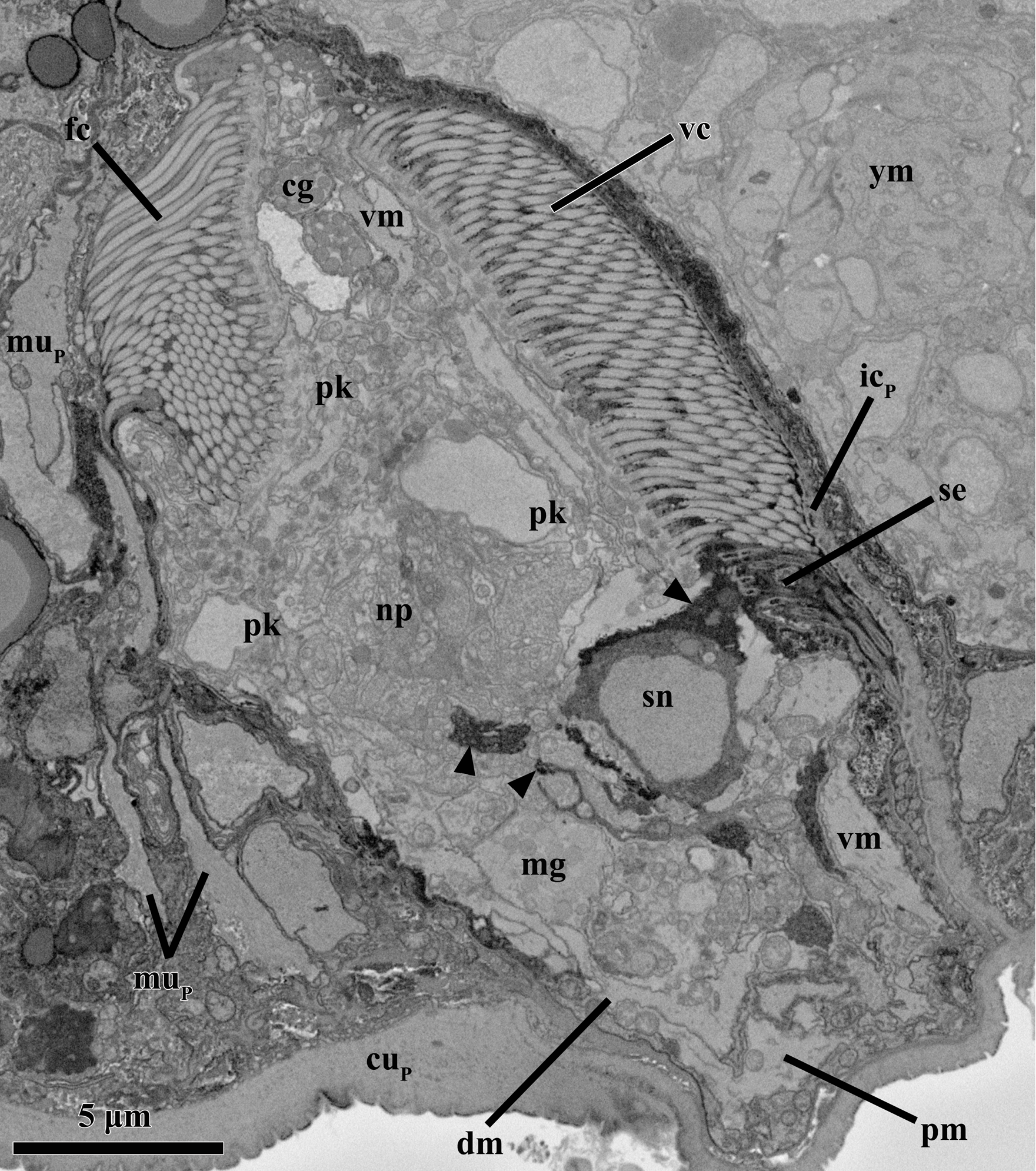
-{Left} Anatomy of the mature male inside the attached Prometheus larva of Symbion pandora.
SEM-SBF micrographs, longitudinal section. A bipolar sensory neuron (sn) projects cell processes (arrowheads) into the neuropil (np) as well as into the sensilla (se) located ventrally. Note the several muscles spanning ventrally (vm), dorsally (dm) and posteriorly (pm). Abbreviations: cg cerebral glands, fc frontal ciliated field, icp internal layer of
larval cuticle, mg medial gland, mup larval muscle fiber, pk perikarya, vc ventral ciliated field, ym young male."
More and extremely detailed information can be found in the original paper on Research Gate
The above material has been selectively sourced and cited as permitted (under the 'Adapt' terms) by the Creative Commons Attribution License under which the paper was distributed. The paper is linked to above and with credits to the originators.
The Youtube video on the right shows Symbion Pandora under an optical microscope. More detailed information can be found here:
https://link.springer.com/article/10.1007/s10152-010-0204-5
Please Note:
Micscape Magazine and Microscopy-uk reproduce this information here stating that it is not the work of our direct contributors. We have produced this document and included these images along with their original links as a 'signpost' for people looking to find out more about this life form.
-{Mol Smith - Microscopy-uk}