Marine Debris from the
Mediterranean Sea
by Brian Darnton, UK
Those who have been hunting for the foraminifera during the
summer break will also have found many other treasures from Davy
Jones' locker: These largely shelly mortalities have been cleaned
by natural means in the sea and sunlight. Very little preparation
is required before they can be dry mounted in a cell for
observation and storage.
The Tube Worms
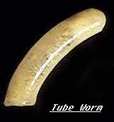
Many worm like creatures build little houses of chalk and
then live in the tubes that they have created. In times of danger
they can withdraw into the protection of the tube.
Some are quite simple and linear or slightly curved, but
others as they grow larger, so the diameter of the tube increases
and a beautiful spiral formation is created, rather like the
fossil Ammonite.
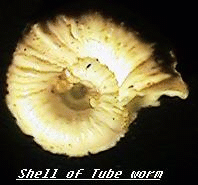
The method of construction is commonly by secretion by the
organism but sometimes a tube is constructed by the selection and
cementing of small plates of sand or even the shells of other
small creatures. One group of these is called the mason worms.
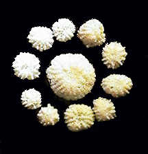
The Sea Mats
These are even more diverse. These are colonial animals that
tend to live within a protective matrix .
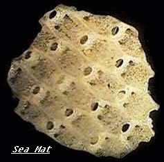
Some appear to be almost woven as an encrustation over rocks
and seaweeds alike. Tentacles are thrust out through apertures
into the surrounding water to feed and breath but at a hint of
danger they are rapidly withdrawn by a strong muscle.
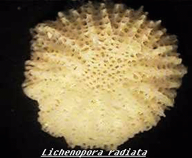
The flat mushroom shaped Lichenopora radiata mimics
the madrepore coral but of course its much smaller. Hundreds of
tube like apertures allow the circular colony to feed and be
protected.
The Sponge Spicules
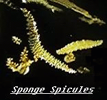 Living sponges are not particularly attractive
in themselves but the spiky reinforcements that support the body
of the sponge in a skeletal like manner can be very ornamental in
crossed polarised light. They too can be found washed up in the
tide mark. Even in British waters they are not at all uncommon.
Living sponges are not particularly attractive
in themselves but the spiky reinforcements that support the body
of the sponge in a skeletal like manner can be very ornamental in
crossed polarised light. They too can be found washed up in the
tide mark. Even in British waters they are not at all uncommon.
The Sea Urchins.
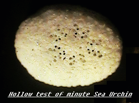
Spines from the urchin family are very common in most parts
of the world. Some are exquisitely fluted like Roman pillars.
Others seem to be cellular. The Victorians used to section the
tubular structure of the spines to reveal a diversity of radial
patterns. Occasionally whole shells can be found of a very small
species. The egg shaped test seems to be able to withstand the
enormous forces of the sea.
Mounting in Cells.
After gluing a thick 19 mm aluminium ring to a slide, a matt
black background can be laid onto the base of the cell using NBS
paint. The shelly remains may be tarnished, in which case two
days immersion in hydrogen peroxide usually cleans them up .
Washing in water through a fine sieve is always a useful
precaution to remove salts.
The dry shells can be glued in the cell using a weakened
solution of gum tragacanth. A small oooo brush may be used for
transfer, to avoid scraping the background. After drying out
under a lamp the coverslip can be applied on a ring of gold size,
to be followed when dry, with an ornamental and protective
external ring of shellac varnish. Do not forget to label the
finished slide.
.
Observation
A low power binocular microscope is ideal for this work .
Reflected illumination from above at 45 degrees should give glare
free clarity of vision.
Comments to the author Brian
Darnton welcomed.
****************
Editor's notes
Brian's other recent articles on marine
subjects for the microscope can be found by typing his surname in
the Library Search index.
Brian prepares and sells a selected range of
strewn and type microscope slides of foraminifera. Visit Brian's Home
Pages for details.
© Microscopy UK or their
contributors.
Published in September 1998
Micscape Magazine.
Please report any Web problems
or offer general comments to the Micscape Editor,
via the contact on current Micscape Index.
Micscape is the on-line monthly
magazine of the Microscopy UK web
site at Microscopy-UK
WIDTH=1
© Onview.net Ltd, Microscopy-UK, and all contributors 1995 onwards. All rights
reserved. Main site is at www.microscopy-uk.org.uk with full mirror at www.microscopy-uk.net.