Click on images to view ca. 150kB
larger images.
Use browser back button to return to article.
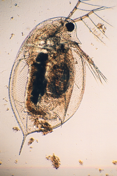 The
colorful views of anisotropic crystals under crossed polars
are a delight to many microscopists. Less well known is the
fact that some types of muscle tissue exhibit similar
properties. These can be well seen in many freshwater
microcrustacea such as the Daphnia pictured here
(right, brightfield).
The
colorful views of anisotropic crystals under crossed polars
are a delight to many microscopists. Less well known is the
fact that some types of muscle tissue exhibit similar
properties. These can be well seen in many freshwater
microcrustacea such as the Daphnia pictured here
(right, brightfield).
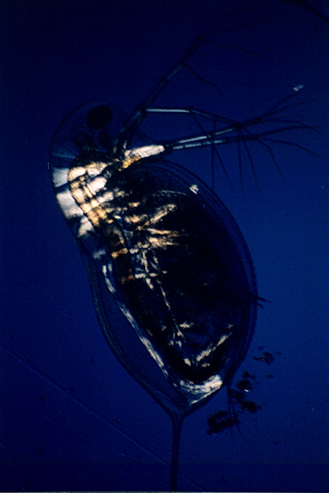
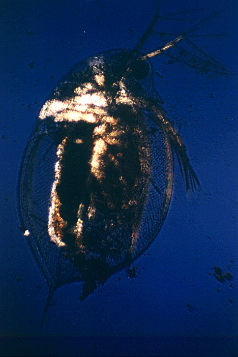 The
crossed polar images (right) very clearly shows the antennal
levator and adductor muscles compared with the brightfield
image. Additional colorful effects (below) can be obtained by
the addition of a wave retarding plate located between the
polarizing filters. The professional ones have very precisely
defined properties. The color effects were obtained here by
simply holding the lid of a plastic petri dish above the
polarizing filter on the illuminator. Scraps of cellophane
from various household sources will work well if of an
appropriate thickness.
The
crossed polar images (right) very clearly shows the antennal
levator and adductor muscles compared with the brightfield
image. Additional colorful effects (below) can be obtained by
the addition of a wave retarding plate located between the
polarizing filters. The professional ones have very precisely
defined properties. The color effects were obtained here by
simply holding the lid of a plastic petri dish above the
polarizing filter on the illuminator. Scraps of cellophane
from various household sources will work well if of an
appropriate thickness.
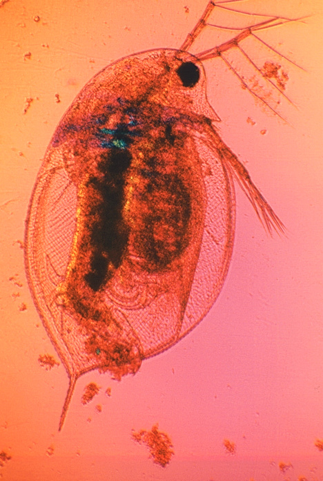 Unlike
crystals, critters can be difficult to keep still long enough
for the exposure times required for crossed polars. The
difference in exposure required for a given subject in both
brightfield and crossed polars can be well beyond the range
of many flash units. These photographs were obtained by using
a microscope slide with two cover slips cemented about 5mm
apart. The subject was placed in a drop of water in the gap,
posed and a cover slip placed on top. The slide was then very
gently heated until the subject was killed and photographed
immediately. I find this system both cheaper and better than
the commercial cavity slides because the depth of the fluid
is even, unlike a hanging drop.
Unlike
crystals, critters can be difficult to keep still long enough
for the exposure times required for crossed polars. The
difference in exposure required for a given subject in both
brightfield and crossed polars can be well beyond the range
of many flash units. These photographs were obtained by using
a microscope slide with two cover slips cemented about 5mm
apart. The subject was placed in a drop of water in the gap,
posed and a cover slip placed on top. The slide was then very
gently heated until the subject was killed and photographed
immediately. I find this system both cheaper and better than
the commercial cavity slides because the depth of the fluid
is even, unlike a hanging drop.