About
the Photography
All the images presented in The Micropolitan Museum were made with the light microscope. The collection was photographed with a Zeiss standard microscope and an old Zeiss photo-microscope. Several methods of illumination were used. Bright field, dark field, phase contrast, differential interference contrast and Rheinberg illumination. Loxophyllum, one of the stars of the Ciliate Center will now demonstrate some of the illuminations used.
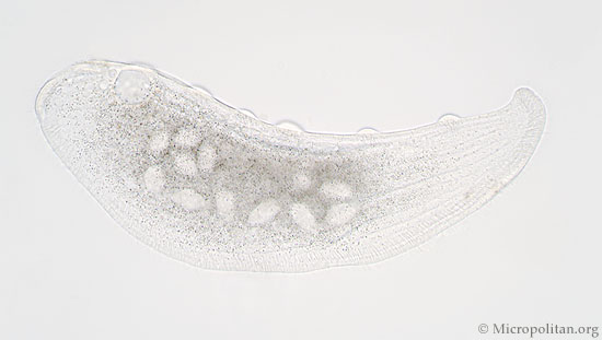
In bright field you can see how transparent this organism is.
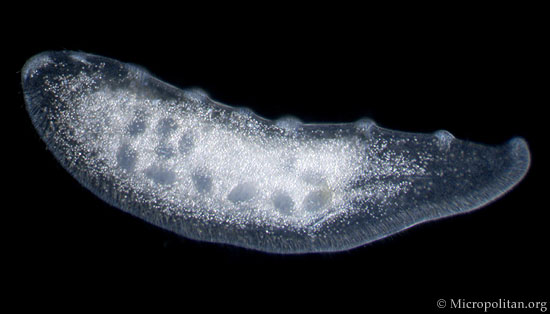
Dark field illuminiation enhances the contrast.
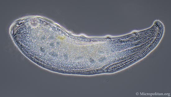
Phase contrast shows more details, like the cilia.
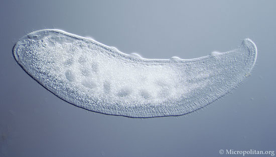
Differential interference contrast creates highlights and shadows.
Many of the images were made on slidefilm (mainly 50 ASA Velvia or 100 ASA Kodak Ektachrome). The slides were scanned and scaled down to 640 pixels wide or 620 pixels high to fit the computer screen. Some sharpening has been added to show a bit more detail in the small images. For the 2008 update of the museum digital photomicrographs were added.
If you like to create good photomicrographs it is not the cost of the equipment that is most important. It is more the manipulative skills of making a good microscope slide. The way you prepare a sample is important for a good result. Apart from some micro-fossils all the organisms presented in the museum were alive when they were photographed. One of the secrets is the use of dots of Vaseline to support the cover-slip. It will prevent damaging the delicate microorganisms. A small talent in hypnosis might also help to slow down these restless microbes.