![]()
Equipment:
Small raw onion
Clean microscope slide
Glass cover slip
A mounted needle (useful)
Tweezers (useful) and fine scissors
Compound microscope with 9X or 10X objective
Optional: Small amount of fountain pen ink (blue or black).
Method:
 Cut a quarter segment from
the onion with a knife (care) and peel off the first scale leaf
(an outer layer which breaks away). Remove one of the inner
layers and bend it back to break it. You should find that when it
breaks the epidermis (thin skin) is still intact. Carefully peel
this away (tweezers are useful for this) and with scissors cut a
small square of this thin skin.
Cut a quarter segment from
the onion with a knife (care) and peel off the first scale leaf
(an outer layer which breaks away). Remove one of the inner
layers and bend it back to break it. You should find that when it
breaks the epidermis (thin skin) is still intact. Carefully peel
this away (tweezers are useful for this) and with scissors cut a
small square of this thin skin.
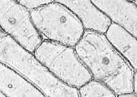
Place the skin on a microscope slide with a drop of water. Cover carefully with a cover slip trying to avoid trapping air bubbles. A mounted needle will make it easier to lower the cover slip onto the slide. Examine under a compound microscope with a 9X-10X objective and you should clearly see the cell structure shown right.
Staining the plant cells (optional): You may wish to try crudely staining the cells to make some features more clearly visible. Before mounting, dip the small piece of onion skin in a few drops of ink, then briefly dip it in clean water, and then prepare the slide as described above. Some cells may take up the blue dye (commonly methlyene blue) found in ink. The nucleus in particular was stained blue when the author tried this with black ink. The proper staining of animal and plant sections requires more involved techniques and proper reagents, but the crude method above suffices for this simple demonstration.
UK amateur microscopists interested in preparing permanent mounts of plant and animal cells can obtain well written booklets describing the techniques together with all the necessary reagents from Northern Biological Supplies (follow Shop links).
Return to Home in Close Up article
Equipment:
Microscope slide
Roll of clear sticky tape (Sellotape or similar)
Compound microscope
 As mentioned in
the main article, human skin cells are regularly replaced and
easily shed. Therefore it is a simple matter to obtain some for
study under the microscope.
As mentioned in
the main article, human skin cells are regularly replaced and
easily shed. Therefore it is a simple matter to obtain some for
study under the microscope.
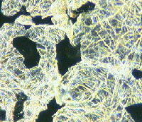
Take an inch or so of the sticky tape and press it firmly down on the back of your hand with your finger nail (to avoid putting fingerprints on the tape). Pull the tape briskly off your hand and mount it sticky side down on the microscope slide, again using the back of a finger nail to press the tape down to remove most air bubbles.
Examine the slide under a compound microscope with a 10X - 20X objective. The general features of the skin should be visible although the author was unable to distinguish individual cells or nuclei when he tried it. The image on the right above shows skin cells under dark field illumination.
Staining the cells (optional): You may wish to
try spreading a small amount of blue fountain pen ink on the back
of your hand first with a cotton bud and allowing to dry. Then
take a skin sample on the inked part of your hand with the tape
as described above. This may stain some of the cells to make them
more clearly visible.
The author Dave Walker, thanks Anne Bruce for describing the
method on which the above preparation is based.
Return to Home in Close Up article
Disclaimer: all the information in this series is given in good faith. However, no responsibility is accepted for damage to property or injury to persons as a result of readers investigating the subjects described. It is up to the reader to judge whether the subjects can be safely viewed in their own home. Younger readers should consult their parents or teacher as appropriate before examining either their own or somebody else's property. Return to top of page