![]()
Introduction to our new series - what you need, who is it aimed at?
Welcome to the third in this bi-monthly series which encourages you to explore in close-up the everyday things around your home.
This month we look more closely at the fabrics you wear everyday, probably without giving them a second glance. We also look at an onion skin to see typical plant cells, and take a sample of human skin to examine animal cells.
Clothes
fabrics
![]()
 Most of us rarely study the intricate and fascinating
way in which fabrics are woven, so why not rummage through your
wardrobe to examine different fabrics. A 10X lens or low power
stereo microscope is required to study them. Some of the fabrics
I found are shown below.
Most of us rarely study the intricate and fascinating
way in which fabrics are woven, so why not rummage through your
wardrobe to examine different fabrics. A 10X lens or low power
stereo microscope is required to study them. Some of the fabrics
I found are shown below.
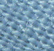
The image on the right is a pale blue shirt (polyester-cotton) at a screen magnification of 20X. The parallel fibres running at right angles to each other are called the warp and weft. It is apparent that each warp and weft strand alternately passes above and below the strand it crosses. This is described as 'plain weave' and is the simplest way of weaving cloth.
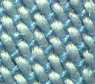 Another pale blue shirt had a
different look and feel, but it was only when I examined it more
closely that I realised why. A close up at the same magnification
is shown on the left. Again it is a 'plain weave' but notice how
it uses both white and blue thread to create the effect. The
white thread is also thicker than the blue.
Another pale blue shirt had a
different look and feel, but it was only when I examined it more
closely that I realised why. A close up at the same magnification
is shown on the left. Again it is a 'plain weave' but notice how
it uses both white and blue thread to create the effect. The
white thread is also thicker than the blue.
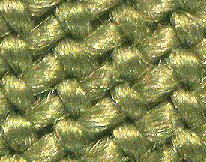 I examined polyester-cotton
trousers which by eye possessed a fine diagonal ribbing. A part
of the beige coloured cloth is shown on the right (screen
magnification 24X). The fine ribbing runs vertically in the
image. By following the path of individual threads it is apparent
that this is not the plain weave used in the shirts shown above,
but instead is a form of weave known as 'twill'.
I examined polyester-cotton
trousers which by eye possessed a fine diagonal ribbing. A part
of the beige coloured cloth is shown on the right (screen
magnification 24X). The fine ribbing runs vertically in the
image. By following the path of individual threads it is apparent
that this is not the plain weave used in the shirts shown above,
but instead is a form of weave known as 'twill'.
In the simplest twill the weft crosses over two
warp yarns, then under one. There are also additional features of
the weave to create the effect seen. Apparently twill weave
drapes better than plain weave and also has a superior strength
for working clothes such as suits, jeans and trousers.
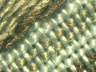 I found a cotton handkerchief
with a chequer pattern on the border and wondered how the pattern
was built up. A close-up of the border is shown on the left and
it is apparent that it is created by a complex use of different
coloured threads and weave patterns.
I found a cotton handkerchief
with a chequer pattern on the border and wondered how the pattern
was built up. A close-up of the border is shown on the left and
it is apparent that it is created by a complex use of different
coloured threads and weave patterns.
So, why not have a look through your own wardrobe ... you only need a 10X hand lens to admire the intricacies of how different fabrics are woven. If there is a lady of the house, the way stockings and tights are woven is particularly interesting.
The properties of the fibres themselves and how man-made and natural fibres look under the microscope is also a fascinating area for study, and we may return to the wardrobe at a later date!
Further reading: See large encyclopaedias using keywords such as textiles and weaving. Eg Encyclopaedia Britannica entry under Industries, Textile. 15th edition, 1993, vol. 21, p579. Encarta 1994 also has an entry under 'Textiles, types of textiles'.
Onion
epidermis - an easy preparation to view plant cells
![]()
 This month we
make one of our first forays into the kitchen. There is a wide
variety of fascinating subjects to study in close-up here, but we
will just study one ... an onion to view an example of plant
cells.
This month we
make one of our first forays into the kitchen. There is a wide
variety of fascinating subjects to study in close-up here, but we
will just study one ... an onion to view an example of plant
cells.
Click here for a simple preparation of an onion epidermis.
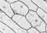
The image on the right shows the cell structure of the epidermis of an onion (screen magnification 100X). The epidermis simply means the thin outer layer. This layer is only one cell thick and therefore the individual cells and the cell membranes can be clearly seen, as well as the nucleus which is the small dark spot in most cells. This cell structure is easy to see with a low power (eg 9X or 10X objective) on a compound microscope.
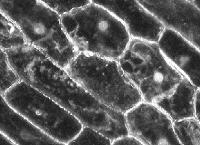 If you have a
dark field illumination stop for the 9-10X objective, a more
dramatic view of the cells can be seen and is shown left. (An
article on dark field illumination and how to make dark field
stops will appear in Micscape shortly).
If you have a
dark field illumination stop for the 9-10X objective, a more
dramatic view of the cells can be seen and is shown left. (An
article on dark field illumination and how to make dark field
stops will appear in Micscape shortly).
Human
skin cells - an easy preparation to view animal cells
![]()
 As we have looked
at the epidermis of a plant it's interesting to compare them with
the epidermal cells from an animal .... you! The cells on your
skin's surface are constantly been replaced and shed, and in fact
most of the dust in your bedroom is skin cells shed overnight
As we have looked
at the epidermis of a plant it's interesting to compare them with
the epidermal cells from an animal .... you! The cells on your
skin's surface are constantly been replaced and shed, and in fact
most of the dust in your bedroom is skin cells shed overnight
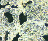
It is very easy to prepare a temporary slide of cells from your own skin. Click here to read how. The image on the right shows a sample of skin cells from the hand (screen magnification 255X) under dark field illumination. It is more difficult to distinguish the individual cells compared with the onion, but the general impression of flat irregular cells is evident. The network of criss-crossing lines is the fine patterning of the skin. Human skin contains keratin which is a fibrous protein which is also the principal constituent of hair.
Incidentally the skin you shed is eaten by
dustmites which live for example in bed mattresses. Micscape has
a range of articles on dustmites.
![]() The sticky tape is also likely
to remove some hairs from the hand and these are also worth
studying. A hair is shown in the image right. The dark irregular
border is an artefact of the preparation as the hair is stuck on
sellotape.
The sticky tape is also likely
to remove some hairs from the hand and these are also worth
studying. A hair is shown in the image right. The dark irregular
border is an artefact of the preparation as the hair is stuck on
sellotape.
Further reading: Most school-level biology textbooks will describe the structure of plant and animal cells and may give simple experiments to study them.
What
do you need? - each subject covered in this series will
have an icon to show what you will need to study an object.
![]() can be viewed with
a 10X hand lens or similar magnifying glass.
can be viewed with
a 10X hand lens or similar magnifying glass.
![]() use a low power stereo
microscope 10X-40X for a clearer look
use a low power stereo
microscope 10X-40X for a clearer look
![]() requires a compound microscope with magnifications 30X
up to 200X
requires a compound microscope with magnifications 30X
up to 200X
Who is the series designed for? We've tried to make it of interest to both the novice and more experienced microscopist. Each topic will have a brief description with images to illustrate the fascinating things you can find to look at. But there will be a click here prompt for many topics which will provide more background information and suggestions for further study.
Disclaimer: all the information in this series is given in good faith. However, no responsibility is accepted for damage to property or injury to persons as a result of readers investigating the subjects described. It is up to the reader to judge whether the subjects can be safely viewed in their own home. Younger readers should consult their parents or teacher as appropriate before examining either their own or somebody else's property. Return to top of page