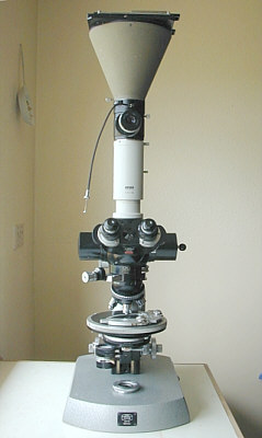
| Carl Zeiss Photo Microscope 1 |
| A Personal Look at a 1950's Photomicroscope
By Paul James (UK) |
 |
 |
| Carl Zeiss Photo Microscope 1 |
| A Personal Look at a 1950's Photomicroscope
By Paul James (UK) |
|
|
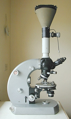
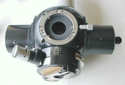
Of the series of Photo-Microscopes that Carl Zeiss designed and built during the middle of the last century, the model illustrated above is the first type ,and is often referred to as the Zeiss Photomic 1. The essential difference between this microscope and most of the others of the day, was that the Photomic had an inbuilt 35mm camera system, as well as being provided with a complex prismatic light pathway slider above the objective changer to allow other photographic possibilities. This particular example sports polarising facilities as well.It is a large microscope by most standards and weighs in at around 42lbs ( 19 kg) excluding accessories such as the heavy 5x4 inch camera ensemble. It probably wouldn't win a gold medal for elegance or style, but as a flexible work horse, with some reservations, it most certainly would.
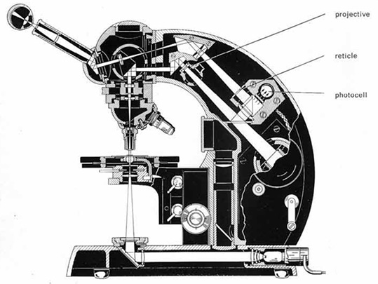
The 'innards' of this rather large stand are complex to say the least and the diagram below shows the basic optical/mechanical internal layout ( There are minor differences among the various models ):-
|
|
Zeiss PMI internal layout .
 |
Zeiss PMIII internal layout for comparison.
The first question we may ask ourselves when seeing this complex array for the first time is probably... "Does any light get back to the eyepiece?" The answer is in the affirmative! Briefly, the light from the objective, when in the 35mm internal camera mode as shown above, passes through a total of about 30 air/glass surfaces between the objective and eyepiece! On paper this sounds very optimistic, but in practise it actually works very well indeed, and all that the observer notices is a reduction in light intensity and no observable change in quality between this mode and the direct route option i.e. all the light from the objective passing to the eyepiece. In fact to my critical eye there appears to be no change of quality in the image as the slider is moved from the internal camera position to the standard 100% light route for normal viewing, which emphasises the design and construction qualities of the internal optics/mechanics.
Internal Camera
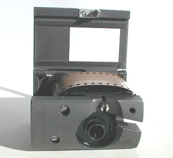 |
 |
| Film tucked in before slack taken up | Mask closed ready to put into body |
 |
 |
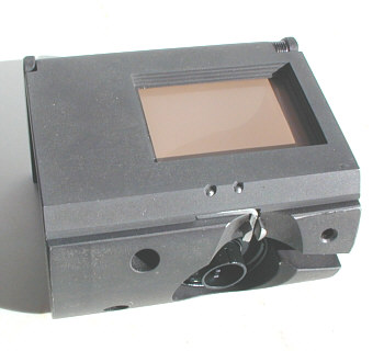
| Film insert now
inside body showing
framing window. |
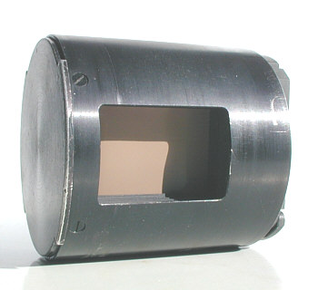
Complete ensemble
ready for insertion into
receptacle in the lower limb. Note that the closed window is automatically opened when inserted into the limb. |
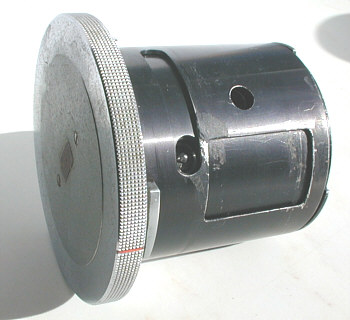
The cylindrical and well designed camera unit is compact and registers neatly into its bayonet seating in the 'scope's body. There's a wind up clockwork film advancing/shutter mechanism which drives the unit either by the depression of a button at the base of the limb, or remotely using an electrical solenoid housed also in the body. The camera has a shutter speed range from Bulb to a 1/250th of a second to encompass most situations that would be encountered, and the system works well. The instrument's eyepiece focussing has to be calibrated so that when the observer views the image, the camera is coincidentally focussed too. This can be a little tedious to set up, but once each eyepiece dioptre setting has been found it can be left, and an occasional 'test' is all that is required to maintain calibration of focus. The object here is that when the instrument is focussed normally for infinity eye accommodation, then so to is the internal camera accommodated or focussed too.I have used film in the Photomic, but hope that a CCD insert of the kind being developed for 35mm SLR's will eventually appear on the market which can be housed inside the camera body, and provide colour digital imagery to the PC.
5x4 Inch Camera
This unit contains an inspection eyepiece housed in a beam splitter, and a shutter together with a substantially made darkslide holder and ground glass screen. It is ideal for photographing still or inert specimens, but since the light from the trinocular port below is not a shared one with the binohead, it is a little inconvenient, and therefore the subject range is somewhat restricted.Nevertheless, it has its uses, and can produce excellent results.
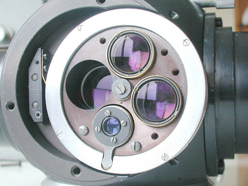
Slide Prism Changer (Light pathway changer)
 |
| Prism slide
changer. Note analyzer on left, bino port centre and
Optovar on right, and objective changer dovetail mounting below. |
This unit contains 4 prism 'units', Giving the following options :-1) Direct vision utilizing 100% of light from objective to bino' head.
2) As above but about 80% of the light directed toward the internal camera, the remaining 20% passes through the bino' head for visual inspection.
3) As above but the image is masked and framed accurately to allow more precise framing for the internal camera, plus the option of increasing magnification up from x 3.2 to x 6 above that of the standard image.
4) 100% of imaging light passes through vertical photo tube as in 'trinocular' configuration.
These individual options are executed by sliding the entire internal prism assembly using a hand operated rod.
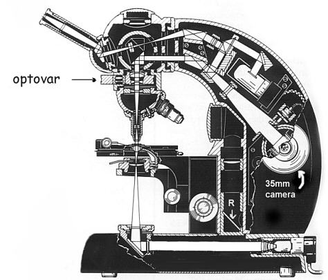
"Optovar"
 |
| Optovar. Note
telescope with focussing tab below, and three
other optics for x1.25, x1.6, x2.0 overall amplification . |
This is Zeiss's own patented rotating magnification unit, mounted directly above the objective changer. Apart from a selection of amplification options of x1.25, x1.6, and x2.0, the Optovar also houses what is in effect a focusing telescope which allows the observer to centre either the phase annuli or the substage/optics. The internal condition of the objectives can be scrutinised as well which is very convenient, but this can also be very alarming too !
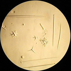 |
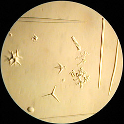 |
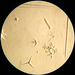 |
| Spicules with x 1.25 Optovar | Same with x 1.6 Optovar | Same with x 2.0 Optovar |
Thus an objective of say x 10 with a x 8 eyepiece ( x80 ), will actually yield images of x 100, x 120 and x 160 with the Optovar. I find this very useful for both photographic and visual purposes, and in combination with the polar rotating stage any specimen can be orientated and amplified to fit into a photo frame with relative ease, without having to move or turn the slide manually.Internal Optics
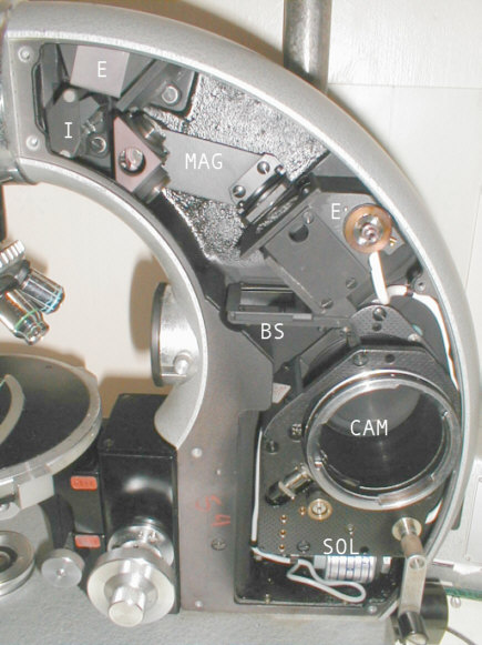 |
The internal optics appear to be very complex, they reveal a well conceived and executed array of mechanics and optics, which is by all accounts quite ambitious, and yet it works well.The light from the objective appears at I and travels through a roof prism and then through an optic in the magnifying changer unit MAG then down through the beam splitter BS finally into the camera housing CAM . The reflected light off the beam splitter carries on at 90 degs into the photo exposure box E and through to the last roof prism E from where it exits into the prism slider and is directed into the binocular head for visual observation. SOL represents the solenoid components for electronic exposure control.
Alignment of all these optical parts is of course vital, and the sample I have appears to be still aligned or collimated fairly well, despite possibly tinkering over the last 45 yrs ! Understandably, the instrument's inherent complexity might put off many would be buyers, especially if the optics have been tampered with and are not collimated correctly, which if professionally aligned might cost a sobering sum?! There are however a number of user adjustments which can if carefully altered in a logical sequence, maintain collimation.
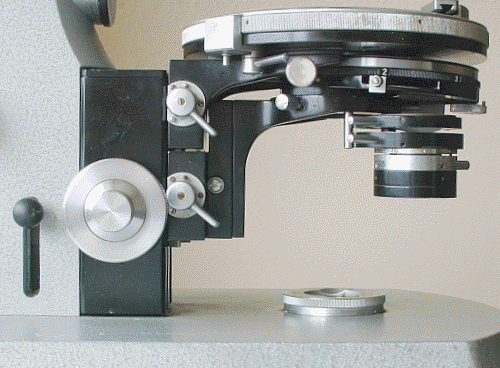
Substage
Below the stage is a phase condenser which is a very nicely designed and thoroughly practical unit, being easily adjustable in all aspects. There are 7 apertures in the Zernicke style rotating disc, 3 for the phase annuli, another containing a remotely controlled iris for brightfield, and 3 spare apertures for darkground etc.. I really like this unit, which also sports a flip-up top element to the Abbe condenser providing up to 0.9 N.A. when necessary.Both the stage and condenser are easily and speedily removable as they have independent locking cams on the " V " slide of the focussing unit upon which they are supported.
 |
Those observers appreciative of plenty of adjustment in the substage will find this 'scope very satisfying and thoroughly practical. The various forms of illumination techniques are easily put into use and there is plenty of scope regarding experimental inserts in the phase unit that this example possesses. One very useful feature I find, is that the iris diaphragm can be shifted laterally by turning the Zernicke disc, and being high up in the substage optics allows very effective oblique illumination in brightfield for trials before using any stops in the filter tray. Though this is not I suspect a deliberate design feature, it is nevertheless a very useful aid to observation being quick and convenient to execute.This iris can also be independently moved in brightfield, so the iris remains collimated with the objective and condenser optics.
Polarising apparatus
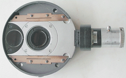 |
This stand sports both a substage 'polariser' and an 'analyser' (as shown above), the latter is housed above the object changer. Note the glass 'window' on the left edge of the unit, which provides dust protection for the internal optics when the unit is withdrawn for normal bright field use etc.This is very convenient, and both units can be moved into the light path when required in a few seconds. Both are well engineered and accurately mounted and are provided with sensitive mechanisms for rotating. There is also a standard circular stage which is calibrated through the 360 degrees and this is also well engineered and very smooth in operation, and held firmly in any position by a cam brake.
Stage
Unfortunately the main stage unit supporting the circular stage etc. has much overhang, which is I think the main cause of its 'flexibility'. My main criticism of this stand is the spongy support of the slides and therefore under high power of around x 250 upwards, any manipulation of the mechanical stage becomes very obvious as the specimen moves in and out of the focal position. This is tedious and causes some time loss when focussing cameras at high power.Trinocular port
This is quite conventional, but houses a very useful collar which supports the 5x4 camera mounting and viewing scope. This can be slide up or down and locked accordingly providing changes of exterior tube length adjustments and eyepiece to camera distances etc. Other cameras can of course be mounted.Personal comments......Pro's & Con's
The ability to photograph with the greatest of ease any specimen in the field of view by simply pressing the release button is very convenient, and requires a little self discipline to prevent excessive use and film wastage!!The substage is excellent and does everything that a substage should be able to do, and in particular the iris diaphragm being located in the Zernicke plate with the phase annuli can be centred too.
Sadly the light pathway changer does not in my particular example house a shared light prism ensemble as is now common in trinocular heads, which means that anyone desiring to use the conventional trinocular port with camera cannot simultaneously see and take exposures at one and the same time, which is a great pity.
The instrument I have, whilst being obviously a serious microscope of intricate design with much diverse capability, does have some flaws. But before launching into any criticism of the instrument, the reader or would be owner should realise that these may be only applicable in my particular case, though I have to stress that it is entirely possible or even likely that other stands of the same designation might well prove to have similar weaknesses ?
My main criticism concerns the fundamental stability and rigidity of the stand, particularly the stage. In use this translates into a stage which lacks rigidity, and when used with powers upwards of about x 250 the normal adjustments of the mechanical stage by hand, disturb focussing in a very obvious way. Also the fine focus when adjusted, sometimes induces very small but discernible alterations of focus when the fingers are removed from the fine focus knobs. In short the high power capabilities of this instrument are somewhat prejudiced. Additional vibration from the release mechanism of the internal camera have not gone unnoticed either, though whether this is after the shutter has opened and closed is not so easy to determine. The photographic results seen using the 35mm internal camera show good images, so perhaps the vibration seen through the oculars is post exposure?
These comments may seem uncharitable, but they are based on simple observation.
The instrument can be used for high power observations of course, but I should make it clear that adjustments of stage and fine focus can become tedious at times, especially when concentrating on very fine detail such as provided by the structure of diatoms during photo sessions.
Rather than castigate out of hand such a complex and very well constructed stand, I have to point out that for very low to medium powers (x 5-200 ), this stand is a great joy to use and I simply wouldn't be without it. The fine focus is, if anything, a wee bit too coarse, but this suits my purposes very well in the low to medium powers, were the conventional fine focus can be too ineffectual. 90% of my interests in microscopy involve the use of low to medium powers, leaving more critical higher power work to a more rigidly compact and conventional stand.
Summary
I have not covered all facets of this 'scope, and in particular have omitted information concerning attachments for reflected light, DIC and any other accessories, as I simply do not have them to hand.I purchased this stand without eyepieces and objectives, but my surplus Wild achromats together with a pair of Spencer x 8 achromatic eyepieces provide exquisite flat field imagery, satisfying my every need. Phase objectives of other makes perform well too. Sadly, Carl Zeiss optics appear to command very high prices in the second hand market, and my experience with other quality makes of 'lower value' suggest that the former are not an essential requirement if a would be purchaser of such a stand were to find it 'bare' too.
All things considered this is a really useful and thoroughly practical stand, and despite potential flaws in stage rigidity, its flexibility, design and quality ensures my continuing usage for hopefully many years to come.
All comments welcome to Paul James.
Related Micscape
resources:
A tour round the Zeiss Photomicroscope III by David Walker,
November 2007
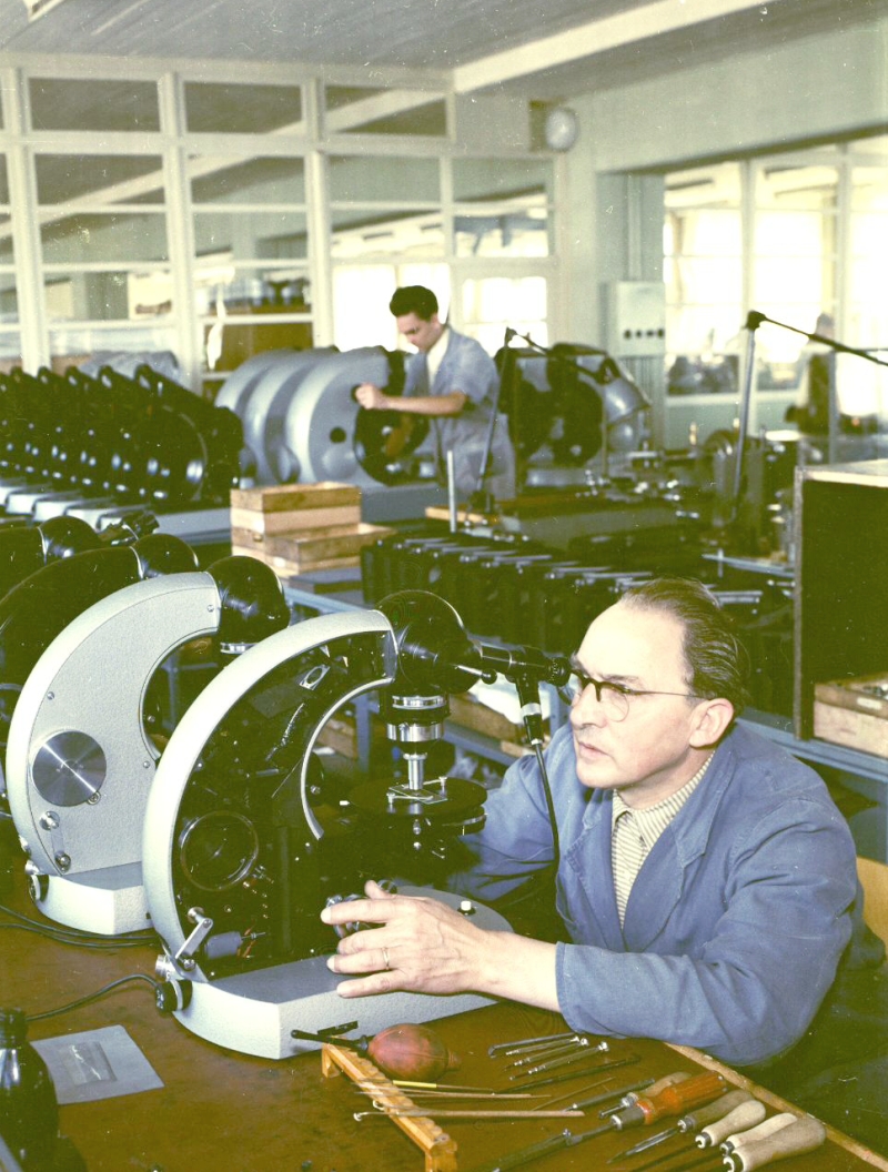
"Production and assembly" in Göttingen in 1957 of the Zeiss Photomicroscope I (the larger Ultraphots are seen in the background).
Image kindly supplied by Dr Johannes Amon, Zeiss and shared with permission.
Please report any Web problems
or offer general comments to the Micscape
Editor,
via the contact on current
Micscape Index.
Micscape is the on-line monthly magazine of the Microscopy UK web site at Microscopy-UK