Comparison of leaf
epidermis in different species
by Anne Bruce
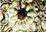
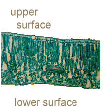 It's quite difficult to visualise that such a thin
structure as a leaf is made up of several layers of cells.
Imagine putting a leaf flat on a surface and cutting across and
then looking end on at the cut edge. The image on the left shows
what you would see under the microscope if you took a very thin
slice from that cut edge. Unfortunately, without access to sharp
blades or a microtome, it can be quite hard to get thin enough
sections to view the cut surface in detail.
It's quite difficult to visualise that such a thin
structure as a leaf is made up of several layers of cells.
Imagine putting a leaf flat on a surface and cutting across and
then looking end on at the cut edge. The image on the left shows
what you would see under the microscope if you took a very thin
slice from that cut edge. Unfortunately, without access to sharp
blades or a microtome, it can be quite hard to get thin enough
sections to view the cut surface in detail.
However, one layer of the leaf, the epidermis,
can be studied quite easily without need or danger of sharp
blades. The epidermis is the outer layer ("epi" -
outside and "dermis" - skin) and is found on the upper
and lower surfaces of the leaf. Nail varnish can be used to make
a "cast" of this surface which can then be viewed under
the microscope.
How to make your cast
- Collect a variety of leaves (trial and
error will determine which sort are the "best"
or "easiest" to cope with)
- Carefully paint a thin layer of nail
varnish over the leaf surface - it is worth while coating
upper and lower epidermis as often there is a difference
between the two
- Leave to dry - again this is down to
trial and error, I have found that leaving the varnish to
dry for a couple of hours, or overnight gives the best
results
- When the varnish is dry, use forceps to
carefully peel it away from the leaf and place on a
slide, preferably with a coverslip over the top
- You are now ready to view under the
microscope
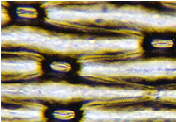
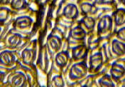
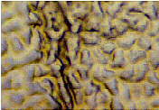
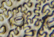
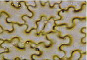
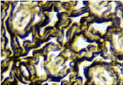
The images below show a variety of different
leaf surfaces and it can be seen that there is not only variation
between species, but also that in certain plants, upper and lower
epidermis differ in stomatal distribution. (You'll find more more
information on the structure and function of stomata in a previous article
in Micscape ).
Comparison of leaf epidermis in different species
|
| |
Spider plant
(Chlorophytum) |
|
 |
 |
 |
Bay leaf (Laurus nobilis)
lower epidermis |
Bay leaf (Laurus nobilis)
upper epidermis |
Morning Glory
(Ipomoea) |
 |
 |
 |
Black Eyed Susan
(Thunbergia alata)
upper epidermis |
Black Eyed Susan
(Thunbergia alata)
lower epidermis |
Black Eyed Susan
(Thunbergia alata)
lower epidermis |
 |
 |
 |
All
images are to the same scale. The width of each of the
images represents approximately 250 microns (ie 0.250mm)
|
A post script
I am grateful to Ron Neumeyer for suggesting
the following tip:
Try laying a tooth pick along the leaf and
include it in the varnishing process. Makes it much easier to
peel off a section.
Comments on the article to the author Anne Bruce
© Microscopy UK or their
contributors.
Please report any Web problems
or offer general comments to the Micscape Editor,
via the contact on current Micscape Index.
Micscape is the on-line monthly
magazine of the Microscopy UK web
site at Microscopy-UK
WIDTH=1
© Onview.net Ltd, Microscopy-UK, and all contributors 1995 onwards. All rights
reserved. Main site is at www.microscopy-uk.org.uk with full mirror at www.microscopy-uk.net.