|
a flatbed scanner by Dave Walker, UK |
The A4 flatbed scanner, in addition to its intended uses, can be regarded as a cheap 'n cheerful digital macroscope for a surprising variety of subjects. Many owners have exploited its potential, as it can be particularly useful for illustrating web articles where the small images don't stretch a scanner's limitations of sharpness and resolution when the subject isn't flat.
Two problems with photomacrography, especially for large subjects, is achieving even lighting and maximising depth of field, the latter which may only be a few mm. This is where the flatbed scanner works well because of the wide even light source and the scanner's intrinsically large depth of field (e.g. 1 - 2cm or more).
Some of the author's previous fun trials using a flatbed scanner are shown in 'Topical Tips 3' (cataloging microscope slides and macroscopic slide subjects) and 'Digital macroscopy in autumn with a flatbed scanner'. Other Micscape contributors are also finding scanners useful, particularly for taking images of microscope slides. For example, see Paul James' 'My favourite slides' in this April 2001 issue and Bill Ells' 'Victorian papered slides'.
As the author's small garden is starting to show some early spring life, I had a stab at scanning some spring plant subjects to see what sort of results can be achieved. (The author's usual route for digital macroscopy is a colour video security camera with 35mm SLR extension tubes and 50mm lens attached and capturing stills with a 'Snappy'.)
Comments to the author are welcomed, email Dave Walker.
Footnote added April 23rd. Many thanks to reader Aydin Örstan who remarks that distortion can occur of 3-D scanned objects, therefore it's advisable to put the object on the centre of the plate to minimise this. Aydin writes 'A good test object to test for distortion is a wine bottle cork placed on its end. If there is no distortion the scanned image should be a circle (provided that the cork is straight). Otherwise, the scanned image will show the circular bottom as well as a portion of the side of the cork.' Aydin also remarks, 'I have scanned 3-D objects as small as 5 mm long & managed to obtain results acceptable for posting on the Internet. An example [of a shell] is at
http://members.aol.com/aydinslibrary/gastrocopta.htm
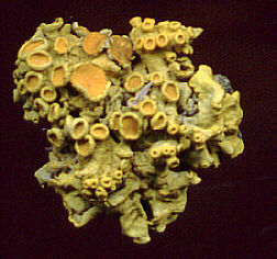
| This small hemispherical
lichen piece (1 cm across) fell off the house roof. The scanner's depth
of field gives quite an acceptable image to show colour and form, albeit
with limited fine detail.
Achieving black textureless backgrounds is easy with a scanner; just leave the lid up! |
 |
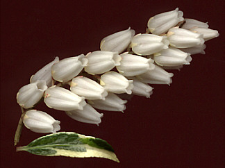
| The scanner's flat lighting (a cold cathode light source in the Plustek) copes well with white subjects without burning out highlights. The sprig is 55mm long and 15mm wide. |
 |
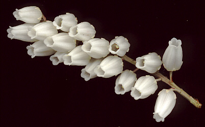
| Another view. The
sprig is just resting on the platter; the subject is 15mm deep.
This is the sort of subject a scanner perhaps works best with, i.e. quite large subjects where form and colour are more important than the finest of detail. For this reason, scans of leaves like ferns can work surprisingly well, (and chilled live dragonflies when expertly handled! - see http://www.dragonflies.org). |
 |
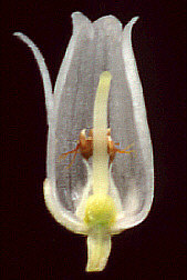
| A single flower (8mm long) from the sprig above, split open. A scanner starts to struggle with fine detail and small subjects which aren't perfectly flat. The image right used a scan resolution of 1200 dpi and resizing with extensive sharpening, but is still a tad soft and lacking in detail. |
 |
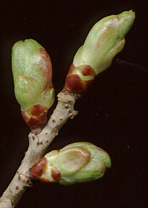
| In spring there is plenty of early plant growth with good macroscopy potential. This is an apple tree twig with buds. |
 |
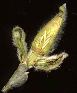
| Cutting the buds open with a sharp knife presents a flatter subject to the scanner, as well as revealing the attractive internal detail. This is a magnolia flower bud. The unripe anthers can be seen. |
 |
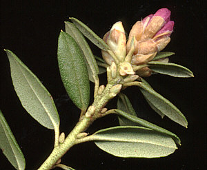
| For leafy twigs like this azalea stem with flower bud, cutting off the leaves in one plane again presents a flatter subject to the scanner. |
 |