My lovely cat that my daughter Melis called her Poessy was only 6 days old when we first saw her. She was trying to grasp one of the nipples of the mother cat. She was one of six newborns and was still blind. Within six weeks she was perfectly able to stand alone and showed a remarkable curiosity about everything around her. When I took Poessy home for the first time to welcome her as the youngest member of my family, she barely filled my hand. Poor little thing!
We arrived home and put her on the floor in the living room. She very carefully nosed the air and walked directly to the cage of our canary. Poessy looked at the bird and remained there for a while, possibly with an intense expectation of a 'First Supper'; but in vain! Such a formidable show of animal instinct was really extraordinary.
Our playful cat grew rapidly and discovered many things around her and in our garden. As I expected, another miraculous action happened, most probably not as a result of her instincts, but because of the influence of her sex hormones. She acquired a companion, a male cat. 'Oh, no!' I said, so far is good, but no further. I couldn't take the responsibility of having another four, perhaps six, new, lovely young cats, oh no. I love my pets but not a pet shop. An appointment was quickly made with the veterinary surgeon before she went into estrus [a cat may go on 'heat' up to 5 times a year!] and she underwent the operation called histero-salpingo-oophorectomy. Sorry for such a long and awful term in Latin but they are almost inevitable in this long article. It simply means that her internal genitalia (also named gonads) are removed. Poessy was thus spayed.
Her ovaries (normally, each female has 2 ovaries!) were oval in shape, satiny in colour and about 10 mm long. The long edge of both were covered by tube-like and undulating structures which finally merged into one larger tube. The Gross anatomy of a cat (and other animals, of course) can easily be found in several, well-illustrated, sites on the Internet. For example, click this to see the gross anatomy of the internal organs: bladder, ovaries, tubes and uterus.
My cat's excised gonads provided me with an excellent opportunity of studying the histology of her ovaries and related tissues. It is also very pleasant to share with you the fascinating microscopic anatomy of a cat's ovary.
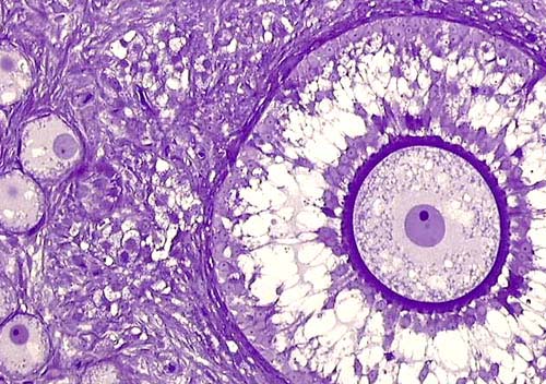
.
Cat's ovary. 2 micrometer (2 µm) thick (actually very thin!) section made after embedding in a plastic matrix. Crystal violet staining. On the lefthand side, we see four primordial egg cells of which two have nuclei and even nucleoli visible as a dark blue dot. The right half of the image is filled by a maturing ovarian follicle in which an egg cell lies at the center. The cytoplasm of the egg cell (ovum) is finely dotted and contains a centrally positioned, purple stained nucleus with an eccentrically situated and blue stained nucleolus. This egg cell is also demarcated by a dark blue and rather thick envelope known as Zona pellucida (which means a transparent area around the cell). Only one sperm can pierce this pellucid zone and then only the head of the sperm penetrates the egg while the tail won't be admitted inside the cell. Once a sperm head and egg cell exchange genetic material it becomes a ONE-celled new individual we call a zygote. A zygote is neither an egg nor a sperm anymore, it is a ONE-celled cat in this presentation (or it may be other animals like humans, sheep, etc.). The illustrated ovum, which is an almost mature egg cell, has a diameter of 160 mm.
Egg cells are also called ovum in singular, ova in plural. When ova mature in the ovary, a tuft of hormone-producing cells develops around them. These cells have the potential of creating higher levels of estrogen and thereafter lutein hormones, and they are under direct hormonal control of the hypophysis. Hypophysis is an appendicular, hormone-secreting organ in the skull and is located in sella turcica, attached to the frontal part of the brain; it functions like the conductor of an orchestra.
A maturing ovum together with its surrounding follicular cells is also called a Graafian follicle. After reaching maturity, a Graafian follicle ruptures into the abdominal cavity and the egg cell is immediately trapped by the fimbrial ends of the Fallopian tubes which, at the same time, transport and harbour the sperm. The meeting of sperm and egg cells takes place there and the fertilization of the egg cells occurs when a sperm enters the ovum. This phenomenon is then followed by two-celled, four-celled, 8-celled, 16-celled (and so on) stages of development of the individual. In other words, it is an embryonic development of a very new, young organism which consists only of undifferentiated cells. This process leads to an embryo which is soon transported to the uterus where it further grows into a fully developed individual by organogenesis (i.e. the formation of the organs during embryonic development). This development occurs during the next 60 days in cats and 9 months or so for humans, and 20 days in mice, etc.
In the following pages, the microscopical details and histological structures of the cat’s ovary will be illustrated.
~~~