The internal, abdominal organs are covered by a serous membrane, hence serosa. The serosa secretes a slightly viscous fluid enabling the smooth gliding of these organs since they constantly move within the abdominal cavity in parallel to their regular functioning. Think about our intestine and large bowel and guess why they should glide from time to time.
The Fallopian tubes must also glide a bit to transport sperm or catch the ova which are expelled from Graafian follicles at the midway of the menstrual cycle. Such rather slow movements are possible in animals thanks to non-striated, smooth-muscle cells. The following images will show us the serosa, the wall of the tubes and uterus and some more structural details of their walls.
It is easy to understand that such complex tissues and organs must be well supplied with blood and requires many blood vessels and capillaries. They also have nerve bundles which form part of the autonomous neural system."
As the name clearly indicates, this system functions harmoniously and requires no conscious effort, unlike the skeletal muscle systems. We cannot order the smooth-muscles of the tube to "Catch the egg cell!" It is just regulated and performed autonomously.
.
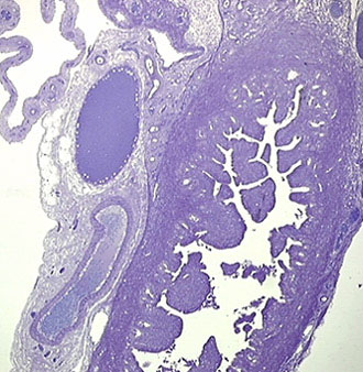
The tubes and especially the uterus (the womb) are rich in glands that secrete a slimy, fluid medium that helps ova passively glide into the uterine cavity. But these organs also have other active mechanisms like vibrating hairs that enforce fertilized ova to move one way. This image illustrates the part of uterus at the site of junction with the tubes. The uterine cavity possesses many finger-like projections in which glands are located. These numerous clefts and projections considerably enlarge the mucous surface. The wall contains bundles of smooth muscle. Outside the muscle layer, there is the serous membrane in which large blood vessels (veins filled with blood cells and empty arterioles) and nerve bundles can be found. All these structures are spread in a loose, connective tissue consisting of collagenous fibrils. As mentioned earlier, the surface of this serous membrane is covered by a serous cell layer.
.
.
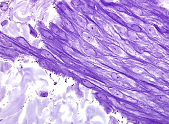
We see here a bundle of smooth muscle cells at successive magnifications, 25x and 40x. Around the bundle, a loosely structured, almost cell-free connective tissue is present.
.
.
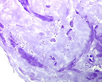
These images illustrate undulating collagen fibrils in that loose connective tissue; 25x and 40x.
~~~