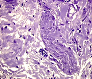
In this moderately cellular connective tissue of the serosa, we see a large nerve bundle on the right. Crystal violet staining. 25x
.
.
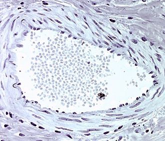
Left: a vein filled with blood cells and showing the smooth muscle cell layer of its wall. 25x. Right: an arteriole with fewer blood cells. Note the spindle-shaped, darkly stained nuclei of the smooth muscle cells of the wall. Toluidine blue staining. 40x.
.
.
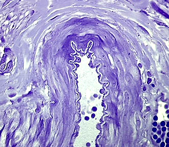
This image of another arteriole clearly shows an elastic, strongly undulating and unstained membrane just beneath the cell layer of the tubular cavity. The arterioles are mainly lined by a layer of flattened endothelial cells also known as intima cells. Can you identify these flat cells in the image, of which a few are still large and visible (look at upper part of the lumen)? Crystal violet staining. 25x
.
.
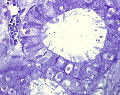
The open end of the Fallopian tube which catches the egg cells. Note that the lining cells show obvious hairy extensions. Both are stained with crystal violet. These hairy structures are constantly moving. This action helps direct the contents of the tubular cavity towards the uterus. Left: 25x. Right: 40x with an 1.6x intermediary projection.
~~~