 .
. 
My Cat's Ovary |
by M. Halit Umar
Page 4 of 6
.
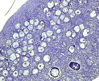
Compared to the Fallopian tubes and uterus, the ovary is a rather compact and firm organ. Although difficult to identify, they are also covered by a cell layer, the stratum germinativum. During embryonic development, this layer produces gonad cells. In these images we see the surface (upper left corner) and groups of primordial egg cells.
In humans, ova are generated once in a lifetime during embryonic development and about 400 of them can ripen, of which only a very limited number give rise to new individuals. Except for some special medical treatments, humans produce only one fully ripe egg cell which is expelled into the abdominal cavity each menstrual cycle, whereas animals like cats are able to produce more than that. This ability frequently results in several kittens in one gestation. This phenomenon is radically different from twins or multiplicities in human pregnancy.
It is thus surprising why Nature chooses at the outset a limited number of ova at the birth of the female, whereas each ejaculation may contain many millions of sperm in the male! And it is also surprising that despite the production of such a huge number of sperms in an ejaculate, only ONE of them can unify with the egg cell! In comparison, production of the sperm is certainly energy consuming and substantially not economic while females represent a higher degree of energy conservation. I suppose that the balance can be made if we think that after fertilization there is still a very long way to go to make a perfect living organism before the birth and as well as during lactation and thereafter.
.
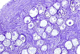
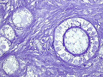
In this higher magnification of the cortical part, we can see the cells lining the ovary which possess groups of ova with prominent nuclei and nucleoli. Hematoxylin stain. 25x with an 1.2 intermediary projection.
.
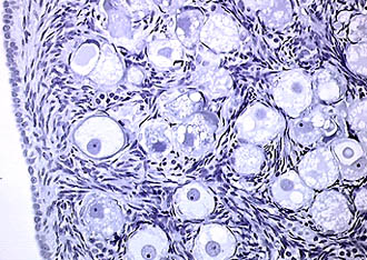
Higher magnification. 40x with 1.6 intermediary projection.
.
 .
. 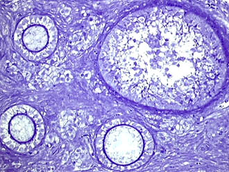
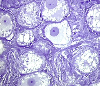
Egg cells are surrounded by densely packed fusiform cells which contribute to follicle development under the influence of hormones. The ovum in the lefthand image is surrounded by a layer of so-called follicle cells. The clear zone (stained blue) developed between follicle cells and egg cell, is the zona pellucida. The righthand image shows a further stage of maturation in the follicle development. The number of follicle cells is strongly increased. 25x
~~~
Published in the April 2001 edition of Micscape Magazine.
Please report any Web problems or offer
general comments to the Micscape
Editor,
via the contact on current Micscape Index.
Micscape is the on-line monthly
magazine of the Microscopy UK web
site at Microscopy-UK