My Cat's Ovary |
by M. Halit Umar
Page 5 of 6
.
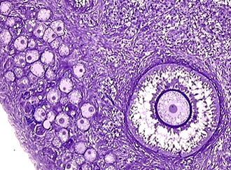
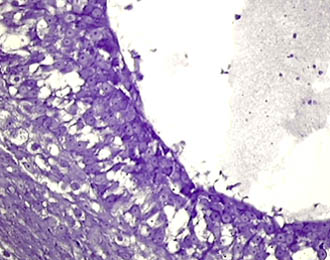
These images show that one egg cell goes into the process of maturation. Note that the follicle cells make a radiating pattern and strongly increased in number. On one edge of the images we clearly observe the surface of the ovary. Crystal violet stain. 25x
.
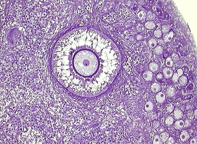
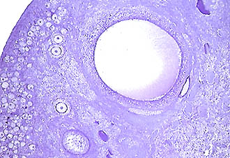
This figure illustrates the apparent multi-layer structure of the follicle cells with an almost clear cytoplasm. Note the zona pellucida and ovarian stroma around this maturing Graafian follicle. Hematoxylin stain. 40x with an intermediary projection of 1.6x.
.
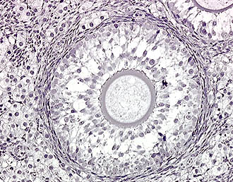
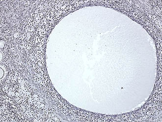
When the maturation of the Graafian follicle is nearly complete, there occurs a local liquefaction within this compact structure followed by formation of a hollow space. Then, the ovum with its prominent zona pellucida and a radiating layer of follicle cells remains on the top of a cell mass, named cumulus oophorus. Crystal violet. 40x
.
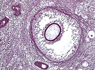
A large amount of fluid accumulate in the follicle together with cell and nuclear rests. Follicle cells become compressed and somewhat flattened by increased tension of the fluid. Soon thereafter, the follicle ruptures into the abdominal cavity and ovum almost immediately captured by the fimbriae of the tubes. Toluidine blue stain. 40x with 1.6x intermediary projection.
.
 .
. 
In case of continuous retention of matured egg cells, the latter degenerate and soon disappear but follicles may remain for a while as liquid filled, large, cystic spherules. 10x.
~~
Published in the April 2001 edition of Micscape Magazine.
Please report any Web problems or offer
general comments to the Micscape
Editor,
via the contact on current Micscape Index.
Micscape is the on-line monthly magazine
of the Microscopy UK web
site at Microscopy-UK