by M. Halit Umar
Page 6 of 6
My Cat's Ovary |
by M. Halit Umar
Page 6 of 6
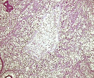
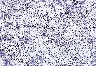
Normally, we expect a visible collapse of the Graafian follicle after discharge of the ovum. The follicle cells become foamy and clear acquiring a yellow tint. This is why we call them corpus luteum, the yellow body. The upper image shows such a yellow body in development. Crystal violet stain. 2.5x. These cells secrete the lutein hormone. The lower image above shows the foamy, clear, luteinized cells of the follicle. Hematoxylin stain. 10x
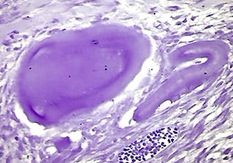
After a while such corpora lutea regress into an almost totally acellular, rubber-like, whitish structures called corpora albicantia. They are actually ovary scar tissue as a reminder of an old site of follicular development. Crystal violet. 2.5x.
These pages show a richly illustrated histological presentation of my cat's ovary. This study helps us to appreciate a biological aspect of Nature and one of the anatomical structures of living beings, in this case of an animal, Felix domesticus, represented by my cat, Poessy.The ovary is a hormonally controlled, dynamic organ and has to cope with cyclic changes during each menstrual cycle. The ovaries in female and testes in male animals are the site of meiotic cell division. By meiosis, the number of chromosomes is halved. So, such gonadic cells are designated as n for the number of their chromosomes. After fertilization, the union of a sperm and egg cell produces immediately an 2n situation. This is the condition in which somatic cells develop, tissues and organs are formed. As soon as an ovary develops, a new reduction process (meiosis) takes place and egg cells are formed. Under normal conditions, no more egg can be formed after the birth of the animal. Contrary to this, in the testes, spermatogonia (stem cells of the sperms) remain active in the meiotic process during the whole sexually active life period. During ageing, sperm production decreases although testes still produce surprisingly huge numbers of them.
Note that the n indicates the haploid condition while 2n the organismal, diploid condition. So, we can easily conclude that animals are moving around all in a diploid state. But do not be surprised that, for example in fungi, almost the whole life span passes in an n state. Only a fungal zygote, for a very short period of time, during sexually reproducing mycelia of mushrooms are in 2n state. This matter is rather complex and not fully understood.
For further reading, the following are links to Britannica's online resources:The ovary in zoology and ovum.
The cat entry explains the main characteristics of the Family Felidae and provides a link to domestic cat resources as well.
Diagram showing the stages of ovum and follicle development.
Biographical information on Reinier de Graaf who first discovered the Graafian follicles.
Please report any Web problems or
offer general comments to the Micscape
Editor,
via the contact on current Micscape
Index.
Micscape is the on-line monthly magazine
of the Microscopy UK web
site at Microscopy-UK